July 2025

Academic Emergency Medicine

None

Annals of Emergency Medicine

Adherence to “Choosing Wisely” Recommendations in Pediatric Emergency Medicine

EMCRIT

EMCrit 403 – What’s the Status of Status – Status Epilepticus 2025 with PulmCrit

Benzodiazepenes

Josh is giving 0.1 mg/kg of Lorazepam (up to 8 mg)

Scott varies by scenario:

Seizing < 5 minutes

Nothing or 2 mg of lorazepam

Status Epilepticus (seizing >5 minutes, but <10 min)

If you have IV access:

Midazolam 5 mg IV

wait 2-3 minutes

Repeat Midazolam 5 mg IV

No IV:

RAMPART dose of 10 mg IM up front

Status Epilepticus (Seizing with EMS prior to arrival, >10 minutes seizing)

Midazolam 10 mg IV or IM (if patient did not get this dose in the field already)

Ketamine

Scott gives Ketamine 1mg/kg with his second dose of midazolam, or immediately following the first 10 mg dose in extended seizures

Ket-Mid Study

PMID [40186980]

Debate with First10EM on this Study

Scheppke Study

PMID: 39642307

Protocol from the study

  • F/u Retrospective Study with comparison group [DOI: 10.1016/j.annemergmed.2024.11.002]
  • Additional support with ketamine as first-line infusion in peds [10.1007/s12028-025-02212-5]

Anti-Epileptics

We both start with Levetiracetam (Keppra) 60 mg/kg (max dose 4.5 g)

Give this whenever it is ready, but do not wait for it to work, if the patient is still seizing, move on to intubation and propofol

ESETT Trial looked at this dosing [10.1056/NEJMoa1905795]

2nd line anti-epileptic:

Lacosamide (Vimpat)

Major contraindications:

  • Pre-existing heart block or conduction system disease.
  • Underlying proarrhythmic conditions (ventricular tachycardia is reported in some cases).
  • Severe hepatic dysfunction

Dose:

400 mg IV over 5 minutes (See IBCC for additional dosing info)

Med QuickRef

able 7. Medications for Status Epilepticus

First-Line Agents

AGENTROUTEDOSAGE
LorazepamIV• 0.1 mg/kg; max 4 mg/dose (some say 8 mg)
• May repeat once
MidazolamIM/IV• Weight 13–40 kg: 5 mg
• Weight >40 kg: 0.2 mg/kg, max 10 mg
• May repeat once
DiazepamPR0.2–0.5 mg/kg, up to 20 mg, 1 time

Second-Line Agents

AGENTROUTEDOSAGE
LevetiracetamIV60 mg/kg; max 4.5 g
Valproate sodiumIV40 mg/kg; max 3 g
FosphenytoinIV20 mg PE/kg; max 1.5 g
PhenobarbitalIV15–20 mg/kg; max 2 g
LacosamideIV200–400 mg

Third-Line Agents

AGENTROUTEDOSAGE
MidazolamInfusionLoad with 0.2 mg/kg and infuse at 0.05–2 mg/kg/hr, with repeat bolus of 0.1–0.2 mg/kg if breakthrough seizure
PropofolInfusionLoad with 1–2 mg/kg and infuse at 30–200 mcg/kg/min
PentobarbitalInfusionLoad with 5–15 mg/kg and infuse at 0.5–5 mg/kg/hr
ThiopentalInfusionLoad with 2–7 mg/kg and infuse at 0.5–5 mg/kg/hr
KetamineInfusionLoad with 0.5–3 mg/kg and infuse at 0.1–5 mg/kg/hr

Meds for Intubation

Scott uses propofol and succinylcholine

Josh uses Ketofol and rocuronium (but lower dose, 0.5-0.6 mg/kg

have a low threshold to intubate these patients!!

General Anesthetic after Intubation

We both use propofol. Scott would consider switching to high dose midazolam drip after 48 hrs if the pt is still seizing.

You need higher doses of propofol to stop seizures, 80-100 mcg/kg/min

These pts will all need norepi, bring it to the bedside immediately after intubation

EMRAP

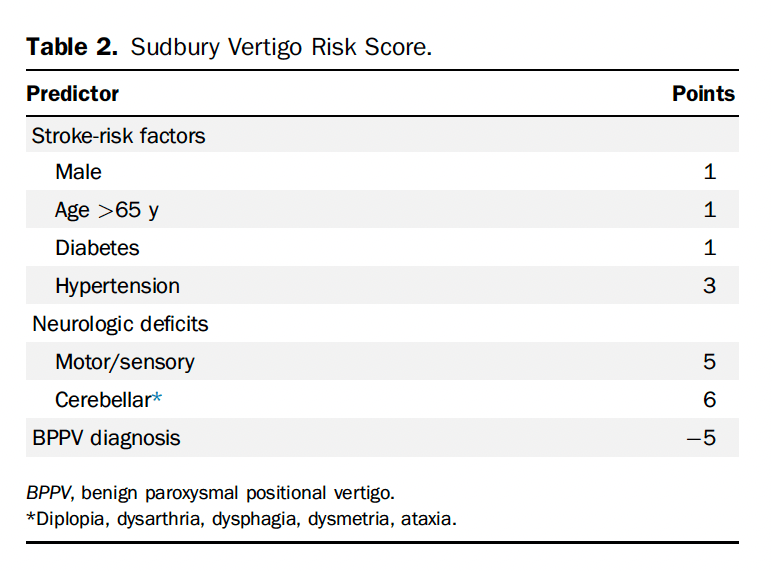
Validating the Sudbury Vertigo Risk Score

  • EDITOR’S COMMENTARY: This study provides the first validation of the recently derived Sudbury Vertigo Score. The score performed very well: scores <5 were associated with 0% risk of serious outcomes, and most patients were in this category. This validation was conducted in a historical cohort of patients at the same 3 sites from which the rule was derived, through retrospective methods leaving much to be desired. I do not think that this rule can be applied to the bedside yet, but it holds promise for helping differentiate this difficult-to-assess patient population.

EMA 2025 June Abstract 6: Risk of Delayed Intubation After Presumed Opioid Overdose in the ED

  • EMA EDITOR’S COMMENTARY: This large prospective study included consecutive patients treated with naloxone for presumed opioid overdose at 10 major toxicology centers in the U.S. Only 1/1,591 patients (0.1%) required an intubation more than 4 hours after arrival, ostensibly because of ongoing toxic drug effects. Although the study has substantial limitations, the findings generally confirm that a 4-hour watch window is sufficient to identify almost all patients who will experience respiratory arrest.
  • EM:RAP HD: Shoulder Reduction by Wrist-Clamping and Shoulder-Lifting

Sickle Cell Disease: Critical Illness

Anand Swaminathan, MD, and Jeff Glassberg MD

Pain control

  • Patients with sickle cell disease frequently present with flare-ups of chronic pain and vaso-occlusive crises.
  • We do not have a good test to determine whether patients are experiencing pain. Our duty is to treat their discomfort.
    • We can look for trends of utilization that may suggest chronic pain.
      • The first step is to treat their pain at this visit.
      • Then we can look for ways to better connect these patients and more appropriately treat the chronic component of this condition.

Transfusion goals:

  • The treatment for sick or ill-appearing sickle cell patients with concern for any type of vaso-occlusive crisis or acute chest is to transfuse.
  • The key to treatment is reducing the fraction of a patient’s own sickled blood to transfused blood.
  • Do not transfuse to a hemoglobin (Hgb) >11 as this increases the risk of stroke.
  • Patients with mild genotypes that have a higher baseline Hgb may need emergent exchange transfusion.

Vaso-occlusive crisis:

  • Create a broad differential diagnosis.
  • Labs: complete blood count ( CBC), reticulocyte count, comprehensive metabolic panel (CMP), lactate dehydrogenase (LDH), Hgb fractionation/plasmapheresis (will help the inpatient team). Assess these labs based on the patient’s baseline if available.
  • Treatment:
    • Oxygen is not beneficial if O2 saturation is >95%.
    • Give a small amount of fluids; eg, 1 L of half-normal saline (1/2NS) or 5% dextrose in half-normal saline (D51/2NS) over several hours.

Acute chest syndrome:

  • Clinically appears like pneumonia (chest pain, cough, hypoxia, infiltrate on CXR)
  • Treat as you would pneumonia (antibiotics, pain control)
  • If they are sick, the key treatment is blood transfusion even if the CXR is normal

Splenic Sequestration:

  • While traditionally a pediatric condition, the early use of hydroxyurea can delay or prevent splenic auto-infarction in children, preserving immunocompetence.
    • Patients who have not auto-infarcted their spleens are at risk for splenic sequestration as an adult.
    • They present with abdominal pain or weakness and can go into hemorrhagic shock.
    • These patients may show massive splenomegaly (>20 cm) on ultrasound.
    • Treatment:
      • Frequent CBC every 4 to 6 hours, frequent transfusion Hgb 1-2 > baseline, and neuro checks every 2 hours.
      • When splenic sequestration reverses, it can lead to significant rise in Hgb, leading to massive strokes; treatment is emergent phlebotomy.

Delayed hemolytic transfusion reaction (DHTR):

  • Patients generate antibodies to both transfused and native blood, leading to an amplified hemolysis of all of their blood.
  • If a patient has received a recent transfusion and presents with an Hgb that is lower than their starting point, this raises concern for DHTR (or bleeding).
  • Treatment includes transfusion and immunosuppression. 

PEARL: The key components to treating sickle cell crisis is to treat the patient’s pain, keep them comfortable, and, if they appear ill from any type of vaso-occlusive crisis, to transfuse to decrease the ratio of sickled to transfused blood.

EMA 2025 June Abstract 13: Nebulized Tranexamic Acid for Post-Tonsillectomy Bleeding

  • EMA EDITOR’S COMMENTARY: If you want to know everything there is to know about the evidence behind nebulized TXA for post-tonsillectomy hemorrhage, this is the article for you. The authors go over all 9 studies on the topic and provide easy-to-follow summaries of each. In sum, although nebulized TXA appears promising as a first-line intervention for PTH, higher-quality prospective studies with standardized protocols are needed before this modality can be universally recommended as standard practice in emergency departments.

EMA 2025 June Abstract 14: Biphasic Anaphylaxis: Incidence and Risk Factors

  • EMA EDITOR’S COMMENTARY: In this single-center study, the authors estimate that biphasic anaphylaxis has an incidence of 16%, occurs an average of 21 hours after the initial reaction, and is generally mild. Emergency providers can use this information to provide anticipatory guidance and return precautions for patients being discharged after successful treatment for anaphylaxis.

Trauma and Transfusion: MTP and More

Jan Shoeneberger, MD, and Kenji Inaba, MD

On the 10-year anniversary of the landmark PROPPR trial, Dr. Kenji Inaba joins Dr. Jan Shoenberger to reflect on its lasting impact on massive transfusion protocols (MTPs). They explore practical challenges in trauma resuscitation, including limited access to O-negative blood, the growing interest in whole blood, and the ethical and logistical considerations clinicians face in real-time decision-making.

1. When to Activate the MTP

  • Clinical triggers:
    • Shock index >1 (HR/SBP)
    • ABC score ≥2 (penetrating trauma, hypotension, positive FAST, tachycardia)
    • Clinical gestalt: patient looks like they’re actively bleeding and unstable
  • Immediate action:
    • Send pink-top tube ASAP for type and cross

2. Standard Transfusion Ratio

  • 1:1:1 ratio = 1 unit packed red blood cells (PRBC): 1 unit fresh frozen plasma (FFP): 1 apheresis platelet unit
    (universal standard as of 2025)

3. Whole Blood

  • Use when available, ideally low-titer O-positive
  • Evidence of benefit exists, but data still limited
  • TROOP trial is ongoing to evaluate whole blood + components vs components alone

4. O-Positive vs O-Negative Emergency Blood Use

  • O-negative preferred for
    • Women <55 years old
    • Children <13 years old
  • O-positive acceptable in life-threatening hemorrhage when O-negative is unavailable
    • Alloimmunization risk is low (<7%)
    • Administer RhoGAM if appropriate

5. Key Practical Reminders

  • Activate MTP early in suspected hemorrhage
  • Always aim for group-specific blood quickly (send pink top)
  • Whole blood can be a first-line option if stocked
  • If only O-positive blood is available, use it in crashing patients (prioritize survival)

PEARL: On this 10-year anniversary of the PROPPR trial, remember to activate MTP early. Always send a pink-top tube to expedite group-specific blood. Use whole blood as the first line if available. In a crashing patient, use O-positive if necessary—survival takes priority.

EMA 2025 June Abstract 15: Vapocoolant Spray vs Lidocaine-Prilocaine for IV Cannulation Pain

  • EMA EDITOR’S COMMENTARY: This relatively straightforward randomized trial indicated no difference in pain reduction during IV placement between the use of a vapocoolant spray and lidocaine-prilocaine cream among adult ED patients. If these treatments really have equal performance, vapocoolant spray is actually the clear winner from a practical perspective, because its immediate onset of action affects procedural time, efficiency, and flow, and the study indicated its higher patient acceptability for future use. The real question is how much added value these treatments provide over using nothing at all; this aspect was not addressed in this study but would influence the ability to recommend routine use.

EMCrit 404 – Facial Trauma

July 19, 2025 by Scott Weingart, MD FCCM 

Look Me in the Face and Tell Me That I Needed to Be Transferred: Defining the Criteria for Transferring Patients with Isolated Facial Injuries [10.1097/TA.0000000000004651]

Bellal Joseph, MD FACS

Dr. Bellal Joseph is the Martin Gluck Endowed Professor of Surgery, Professor of Neurosurgery, Chief of General Surgery and Chief Division of Trauma, Critical Care, Burns & Emergency Surgery, at the University of Arizona. He is also the Vice Chair of Research for the Department of Surgery.

Imaging

Who are you getting specific Face CTs on?

  • Anyone with external signs of trauma or with facial tenderness
  • Scott also gets it on any super sick patient

Who are you getting temporal cuts on?

  • Anyone with a temporal bone fracture

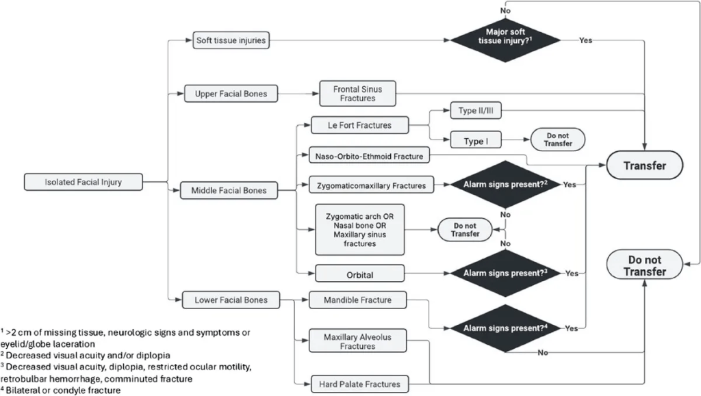
Transfer of Facial Trauma (Facial Injury Guidelines)

Upper Facial Bone Fxs

Frontal Sinus Fractures

  • All should get transferred
  • Look for cerebrospinal fluid (CSF) rhinorrhea
  • All are going to get either observation or operation

Middle Facial Bone Fxs

Radiology Tip: if there is no opacification of the maxillary sinuses, then the only fracture you will miss is a nasal bone fracture

Orbital Fractures

Transfer if decreased visual acuity, diplopia, restricted ocular motility, retrobulbar hemorrhage, or comminuted fracture

Zygomatic Arch, Nasal Bone, Maxillary Sinus Fractures

May need drainage of septal hematoma

Easier to reduce nasal fractures when swelling goes down

Alveolar fracture doesn’t preclude discharge

Zygomaticomaxillary Complex Fractures

Here the complex refers to the components of the fracture pattern, not the complexity

aka quadripod fracture or quadramalar fracture

Transfer if diplopia or decreased visual acuity (On the show, we agreed, decreased ocular motility as well)

Transfer Naso-Orbito-Ethmoid Fractures (NOE) these fxs connote a high mechanism injury

Le Fort Fractures

Type I – Do not transfer

Type II/III – transfer

Lower Facial Bone Fxs

Maxillary Alveolar and Hard Palate Fractures

Do not transfer

Speak with consultants for malocclusion–depends on ability to get the pt in for follow-up

Mandibular Fractures

Transfer bilateral mandibular fractures or condylar fractures

Soft Tissue Injuries

Transfer if >2cm of flap defect or missing tissue, neurologic signs/symptoms, eyelid or globe laceration

Neuro signs and symptoms include loss of sensation, paresthesia, and facial muscle weakness

Additional Tips

Remember to Check the Hearing

Some of these fractures and penetrating injuries can affect the auditory apparatus

Midface Hemorrhage

First10 EM

None

NEJM

Self-Neglect in Older People — A Clinical, Social, and Ethical Dilemma

  • Consider consulting APS for patients with self-neglect that don’t qualify for admission.

Tirzepatide as Compared with Semaglutide for the Treatment of Obesity

  • Tirzepatide (Zepbound) more effective than Semglutide (Ozempic/Wegovy)

Intravenous Tenecteplase before Thrombectomy in Stroke

  • Patients with CVA<4.5hrs did better with lytics before thrombectomy than thrombectomy without lyrics.

REBELEM

MDCalc Wars: Stop Before the CT! — Are You Using PERC or Wells Correctly

June 2025

Academic Emergency Medicine

Clinical decision instruments for predicting mortality in patients with cirrhosis seeking emergency department care

  • MELD 3.0 available on MDCALC if you search for MELD

Annals of Emergency Medicine

Risk of Delayed Intubation After Presumed Opioid Overdose in the Emergency Department

  • Only 0.6% of patients with opioid overdose
  • Of the 1,591 patients included, only 9 (0.6%) required delayed intubation. Eight of these patients had nonrespiratory-related conditions contributing to the need for intubation. One patient only had respiratory-related conditions, had respiratory acidosis, and received a total of 6.4 mg naloxone before intubation.

Severity Indices of Diquat Poisoning for Triage and Prognosis in Acute Diquat Poisoning: A Multicenter Prospective Cohort Study

EMCRIT

EMCrit RACC-Lit Review – June 2025

  • Shock index identifies compensated shock in the ‘Normotensive’ trauma patient
  • Patients with SBP > 90 mmHg on arrival were identified from our trauma center registry. SI was calculated by arrival heart rate divided by arrival SBP. Patients were stratified by SI using the following thresholds: ≤ 0.7,
    • > 0.7 to 0.9 odds ratio to receive blood within 1 hour: 2.5
    • > 0.9 to 1.1 8.2
    • > 1.1 to 1.3 24.9
    • > 1.3 59

Peri-Intubation Adverse Events and Clinical Outcomes in Emergency Department Patients: The BARCO Study. Maia, Ian Ward A., Bruno A. M. Pinheiro Besen, Lucas Oliveira J. E. Silva, Rafael von Hellmann, Ludhmila Abrahao Hajjar, Benjamin J. Sandefur, Daniel Fontana Pedrollo, et al. Critical Care (London, England) 29, no. 1 (April 17, 2025):

  • Among 2846 patients, major adverse events occurred in 919 (32.3%) intubations, most frequently new hemodynamic instability (20.0%), followed by severe hypoxemia (12.5%) and cardiac arrest (3.5%). The overall 28-day mortality was 45.1%. Patients experiencing any major adverse event had a significantly higher 28-day mortality (57.6 vs 39.2%; aHR 1.43, 95% CI 1.26–1.62; p < 0.001). Sensitivity analyses confirmed these findings. Successful first-attempt intubation was associated with a reduced likelihood of major adverse events (aOR 0.52; 95% CI 0.41–0.65; p < 0.001).

Optimal Head-of-Bed Positioning Before Thrombectomy in Large Vessel Occlusion Stroke: A Randomized Clinical Trial.

  •  Results suggest that 0° head positioning for patients with acute LVO was a protective maneuver to maintain clinical stability in the prethrombectomy phase while awaiting definitive treatment.

EMCrit Guest Post – In Defense of Dignity by Chris Hicks, MD

  • Cites last month’s post Mind of the Resuscitationist 4 goals of the resuscitationist:
    • Save Lives/Curative Care
    • Relieve Suffering
    • Enhance Dignity
    • Find Connection
  • Deep dive on enhancing dignity
    • 1. Stop talking around your patients
    • 2. Constant updates
    • 3. Say their name
    • 4. Shared decision making

EMCrit 402 – Sea Change on Salt Water (More Rapid Sodium Normalization in Hyponatremia)? with KidneyBoy

  • New Systematic Review found that patient’s had worse outcomes with slow correction
    • Comparative studies assessing
      • rapid (≥8-10 mEq/L per 24 hours) vs
      • slow (<8 or 6-10 mEq/L per 24 hours) and
      • very slow (<4-6 mEq/L per 24 hours) correction of severe hyponatremia (serum sodium <120 mEq/L or <125 mEq/L plus severe symptoms) in hospitalized patients.
    • Slow correction and very slow correction of severe hyponatremia were associated with an increased risk of mortality and hospital LOS compared to rapid correction.
    • Rapid correction was not associated with a statistically significant increased risk of ODS.
  • 2 Approaches offered in the post:

OR

Pediatric Tracheotomy

Needles fail, use a knife.

EMCrit Wee – Ketamine for Early Status Epilepticus Debate with First10EM

EMRAP

Diabetes in Your Feeties

Whitney Johnson, MD, and Evan Laveman, MD

Treatment

  • Do not give antibiotics if there is no erythema, local swelling, or signs of inflammation. 
  • Diabetic wounds can be chronic and can take a long time to heal.
    • These patients respond well to long-term wound care.
  • >50% of diabetic wounds that do appear to be infected are caused by Staphylococcus.
  • The bioavailability of PO vs IV antibiotics is similar, and many DFIs can be treated with PO antibiotics.
    • Mild infections: 7-14 day course of antibiotics
    • Osteomyelitis: 4-6 weeks of antibiotics

Dressings

  • Wet to dry dressing (once-a-day dressing change)
  • Xeroform dressing with overlying gauze
    • Gentle wrap (not tight or compressive wrapping)
  • Do not use non-adherent dressing or leave wound open
  • Do not use hydrogen peroxide on wound

Wound Care Centers

  • Wound care centers provide management and long-term treatment of surgical wounds, diabetic foot infections, and chronic wounds.
  • DFIs can take months to a year to heal completely.
    • Wound care centers are a great resource for patients: someone is familiar with their wounds and knowledgeable about the entire course of treatment.
  • Wound care centers can manage footwear, order orthotics and do vascular assessments, assess need for lower extremity stents, manage lower extremity edema, and speak with orthopedic surgery and vascular surgery.

Novel Therapies

  • Hyperbaric oxygen therapy for DFIs
    • Breathing in compressed oxygen in a compressed environment
    • Taking place across the lungs and hyperoxygenating the blood
  • Hyperbaric oxygen therapy is helpful for DFIs, wound healing, and osteomyelitis because these areas can have poor microvascular circulation and oxygen tension.
    • Poor microvascular circulation = poor oxygen-mediated immune response, poor effectiveness of antibiotics, and poor wound healing.
  • Generally, 4 weeks of aggressive wound care is required before considering hyperbaric therapy.
    • It is used in cases where wounds are taking much longer to heal than anticipated or where there are elements of chronic/refractory osteomyelitis.

Disposition

  • A patient who is not systemically ill and does not have any signs of severe peripheral arterial diseases can be discharged home:
    • Palpable pulses, capillary refill ❤ seconds, no evidence of sausage digit swelling
    • Can follow-up within 1-2 weeks with primary care or wound care physician
      • Patients who do not have good outpatient follow-up can return to ED 2 weeks after antibiotic course for wound recheck.
  • Consider referring patients to a wound care center or wound care physician.
  • Look at the patient’s footwear.

PEARL: Look for signs of acute infection on top of chronic diabetic foot wounds. High-risk features include systemic signs of infection (tachycardia, fever) and/or erythema, edema (> 2 cm beyond wound), or warmth. Consider referring patients to a wound care center or wound care physician. 

Hypokalemia

Anand Swaminathan, MD, and George Willis, MD

Dr. Anand Swaminathan and Dr. George Willis take a deep dive on hypokalemia. Their conversation will take your management of hypokalemia to the next level. Part 1 covers the clinical presentation of hypokalemia and potassium repletion.

  • Clinical Presentation
    • Patients may complain of fatigue or feeling run down.
    • Cardiac effects include dysrhythmias, including torsades.
    • There may be neuromuscular weakness and, in extreme cases, paralysis or rhabdomyolysis. 
  • Potassium Repletion
    • There is no significant difference in efficacy between different forms of potassium:
      • May administer potassium bicarbonate or potassium citrate in the acidotic patient
      • May opt to avoid potassium chloride in the hyperchloremic patient
      • May administer potassium phosphate in the hypophosphatemic patient
    • 10 mEq of potassium repletion results in a 0.1 mEq/L change in serum potassium level.
      • Potassium levels <3.0 mEq/L will require much larger amounts of repletion to affect serum levels.
    • Oral repletion
      • Preferred method of repletion
      • Best suited for mild hypokalemia (>3.0 mEq/L) 
      • Can give 40-60 mEq/hour 
    • Peripheral IV lines
      • Best suited for PO-intolerant patients or levels <3.0 mEq/L
      • Maximum rate of 10 mEq/hour to avoid painful irritation to small veins
      • Can achieve faster rates by giving through multiple IVs or mixed into IV fluids
    • Central venous lines
      • Reserved for profound hypokalemia and unstable patients
      • As fast as 80 mEq/hour 
      • Patients should be on continuous cardiac monitoring 
    • For patients in cardiac arrest secondary to hypokalemia, you can push 40 mEq of potassium chloride through a peripheral IV.
    • Recheck potassium levels at least 1 hour after IV repletion to allow time for intracellular shifts.
  • Magnesium Repletion
    • Hypomagnesemia results in renal potassium wasting.
    • IV, rather than oral, magnesium repletion is required for hypomagnesemic patients.
    • Magnesium levels of 1.0 or less:
      • Give 4 g of magnesium prior to potassium repletion
    • Magnesium levels of 1.5 or higher:
      • Give 1-2 g of magnesium prior to potassium repletion
  • Disposition
    • ICU indications
      • Patients with neuromuscular complaints – require monitoring for diaphragmatic involvement and respiratory compromise
      • Severe ECG findings, including non-sustained ventricular tachycardia, multiple premature ventricular contractions (PVCs), torsades, or prolonged intervals
      • Level-specific hospital policy
    • Admission
      • Patients with levels <2.8 mEq/L will require ~30 mEq to cause 0.1 mEq/L change in serum potassium level
      • Moderate to severe cases where a cause has not been identified
    • Discharge home
      • Patients with levels >3.0 mEq/L and mild symptoms can be discharged without repeat level after repletion.
      • Patients with levels of 2.8 to 3.0 mEq/L can be discharged after repletion and a repeat level.
      • If patients are taking thiazide or loop diuretics, consider starting low-dose potassium supplementation with close follow-up
      • Counsel patients regarding high-potassium foods (eg, banana, orange juice, sweet potato, avocado).

IV Antibiotics Aren’t Special

Justin Morgenstern, MD

Is the PO (oral) route the way to go? Intravenous (IV) antibiotics are NOT superior to oral antibiotics and show identical treatment efficacy in multiple studies for different types of infection.

Introduction

  • The antibiotic (despite route of administration: PO or IV) is an identical molecule, no matter how it got into the body.
  • The only thing that matters is the concentration of that molecule and how much actually makes it into the tissues (bioavailability).
    • If there is a pill formulation of an antibiotic, it will have decent bioavailability. 
    • The oral route usually takes 30 minutes longer to absorb compared with IV.
    • For oral antibiotics to work, the patient needs to have a functional gastrointestinal (GI) tract.
      • IV is a better route for patients who may be hypoperfusing their GI tract (eg, critically ill or vomiting patient).
  • To date, there are no articles on PubMed that demonstrate that IV antibiotics are superior to oral antibiotics. 

Pyelonephritis 

  • 2014 Cochrane Review
    • 27 randomized controlled trials (RCTs) with 4,500 patients across a range of outcomes:
      • No differences between IV and oral 
  • 2007 Cochrane Review
    • 15 RCTs with both adult and children with severe urinary tract infection (UTI)/pyelonephritis:
      • No evidence that oral antibiotics are less effective than IV
  • An RCT in pregnant women with pyelonephritis (excluded patients with bacteremia):
    • IV and oral antibiotics had identical outcomes 
  • An RCT that compared IV and oral ciprofloxacin for hospitalized patients with severe pyelonephritis:
    • Outcomes were identical 

Pneumonia

  • Multicenter RCT of 1,700 children comparing IV penicillin to amoxicillin:
    • Treatment failure was identical
  • Multicenter RCT with more than 2,000 children comparing IV ampicillin to oral amoxicillin:
    • Oral antibiotics were just as good as IV
  • Multicenter RCT with only 100 adult patients:
    • Oral fluoroquinolone is equal to IV cefotaxime
  • Single-center RCT with 85 hospitalized patient with non-severe pneumonia were randomized to receive IV versus oral antibiotics:
    • No difference between IV and oral

Other Types of Infections

  • There are good data for endocarditis, febrile neutropenia, and acute osteomyelitis, showing that oral antibiotics are as good as IV antibiotics. 
  • The OVIVA trial had more than 1,000 patients with complex orthopedic infections:
    • Septic arthritis, prosthetic joint infections, vertebral osteomyelitis, and discitis 
    • Patients needed 6 weeks of antibiotics (randomized to receive oral vs IV) and outcomes were identical

Oral vs IV Antibiotics in the Hospital

  • Oral antibiotics promote movement and ambulation, whereas IVs can keep patients bed bound.
  • IV supplies are more expensive than pills. 
  • IV placement and maintenance is more time consuming for nursing staff.
  • IVs have high risk for infection, clotting, and patient harm.

PEARL: IV antibiotics are NOT superior to oral antibiotics. Take into consideration when IV antibiotics may be the better treatment choice (eg, for critically ill patients or patients who cannot tolerate PO or are vomiting). 

First10 EM

None

NEJM

None

REBELEM

REBEL Core Cast 138.0: A Simple Bedside Approach to Shock

  • Narrow Pulse Pressure? Cardiogenic, hypovolemic, or obstructive shock 
  • Wide Pulse Pressure? Distributive shock 

REBEL Core Cast 134.0 – Acetaminophen Toxicity

Definition and Physiology

  • After ingestion of a therapeutic dose, immediate release APAP is absorbed with a time to peak concentration anywhere between 30-45 minutes. In the context of extended-release, formulations, full absorption is typically reached by 4 hours post-ingestion.1
  • In therapeutic dosing, the vast majority of APAP undergoes hepatic conjugation with glucuronide or sulfate to form benign metabolites that ultimately get excreted in the urine. The remaining ~5% is oxidized by CYP2E1 to form N-acetyl-p-benzoquinoeimine (NAPQI). NAPQI is hepatotoxic. Glutathione combines with NAPQI to generate non-toxic metabolites that are also eliminated in the urine.

Clinical Manifestations and Diagnostic Evaluation

  • The clinical course of acute APAP toxicity is classically broken into four different stages.
    • Stage1: this is generally within 24 hours. Patients are either asymptomatic or have non-specific GI symptoms (nausea, vomiting, malaise). At this point, hepatic function testing is normal.
    • Stage2: ~24-72 hours. The onset of hepatic injury marks this stage. Aspartate aminotransferase (AST) is the most sensitive marker to detect hepatic dysfunction; AST elevated is nearly universal by 36 hours post-ingestion.
    • Stage3: defined as peak hepatotoxicity; generally between 72-96 hours post-ingestion. Patients may manifest hepatic encephalopathy or coma. AST and/or ALT might rise above 10,000 IU/L. Other lab abnormalities include: INR/PT, glucose, lactate, pH, and creatinine. Death from fulminant hepatic failure usually occurs anywhere between 3-5 days after an acute ingestion. Mortality is often secondary to multiorgan failure, ARDS, sepsis, or cerebral edema.
    • Stage4: often called the “recovery phase.” Patient who survive demonstrate complete hepatic generation without any evidence of hepatic dysfunction.
  • The following labs should be obtained for severe APAP ingestions:
    • APAP Concentration, hepatic panel, pH, coagulation panel, renal function, lactate and phosphate. These labs will ultimately dictate disposition (see King’s College Criteria below)

Management

  • Consider GI decontamination with activated charcoal as this can reduce systemic absorption and limit subsequent clinical sequalae.
  • Ingestions should be classified as acute or repeated supratherapeutic (“chronic” ingestions)
    • Single Acute Ingestion
      • If feasible, obtain a 4 hour post-ingestion APAP concentration. Any concentration earlier than 4 hours is uninterpretable as subsequent concentrations may increase or decrease depending on the clinical scenario.
      • Concentrations between 4-8 hour post-ingestion can be plotted on the Rumack-Matthew nomogram to determine when NAC should be initiated.
      • If the APAP concentration is above the plotted line, NAC should be started.
        • NAC is nearly 100% effective if started within 8 hours post-ingestion.2
      • If an APAP concentration is unable to be drawn before 8 hours or if LFTs are already elevated, NAC should be empirically started if the pre-test probability is high enough for clinical concern.
    • Repeated Supratherapeutic/Chronic Ingestions
      • Cannot apply the Rumack-Matthew Nomogram
      • If LFTs are elevated or if there is a positive APAP concentration, NAC should generally be started however consultation with a toxicologist or Poison Control Center is advised as these cases are often complicated.
  • N-Acetyl-Cysteine (NAC)Dosing
    • “3 Bag Protocol” – 21 hour regimen
      • 150mg/kg over 1 hour loading dose
      • 50mg/kg over 4 hours = 12.5 mg/kg/hr
      • 100mg/kg over 16 hours = 6.25 mg/kg/hr
    • Risk: anaphylactoid reaction
      • Reaction is rate related and typically occurs during the loading dose
      • Symptoms: flushing, urticaria.
    • NAC should be continued until all of the following criteria are met:
      • Negative APAP concentration
      • “Significant Decreased in AST”: defined as either <1000 IU/L or a 25-50% drop from the peak.
      • No evidence of hepatic failure
    • If criteria are not met, the third bag should be extended indefinitely.
  • The King’s College Criteria should be used as this set of lab work is used to determine which patients should be referred for possible liver transplant evaluation.3, 4

Take Home Points

  1. In overdose, acetaminophen leads to generation of NAPQI which is hepatotoxic. N-Acetylcysteine (NAC) is the antidote of choice and ideally should be administered within 8 hours of an acute ingestion.
  2. To determine which patients should be treated with antidotal therapy, the Rumack-Matthew Nomogram should be utilized. Of note, this nomogram was validated for a single concentration obtained at or greater than 4 hours after a single, acute ingestion. (i.e. patients with repeated ingestions cannot be applied to the nomogram).
  3. In patients with a high pre-test probability of APAP poisoning, the King’s College Criteria should be considered; this is a set of lab markers that help determine when patients should be immediately referred for liver transplant.
  4. While physiologic plausibility exists for the use of fomepizole to treat severe APAP toxicity, no large scale human studies exist at this time to suggest that it should be routinely given for toxicity. As with all cases of toxicity, please call your local poison control center for assistance.

Combination Inhalers in Asthma: Time to Switch?

  • Prescribe comb inhalers (LABA/ICS) for rescue therapy to patients with asthma in addition to their maintenance medications. The inhaler of choice may vary based on cost and availability.

May 2025

Academic Emergency Medicine

Utility of predictive scales for emergency department patients
with a new diagnosis of aneurysmal subarachnoid hemorrhage

  • The Hunt and Hess Scale (HHS) assesses headache severity, neck stiffness, neurological deficits, and consciousness level, with higher scores correlating with increased mortality.7 However, its subjectivity and lack of clarity have drawn criticism.
  • The World Federation of Neurological Surgeons Scale (WFNS), incorporating the Glasgow Coma Scale (GCS) and assessments for hemiparesis or aphasia, is more objective and has demonstrated stronger predictive capabilities.8

Emergency department–initiated oral naltrexone for patients with moderate to severe alcohol use disorder: A pilot feasibility study

  • ED-initiated oral naltrexone is feasible and acceptable for patients with moderate to severe AUD. While engagement in treatment was moderate, significant reductions in alcohol craving and improvements in quality of life suggest potential benefits. Further research is warranted to confirm these findings.
  • California Bridge Program recommendations:
    • Gabapentin 600-900 TID for alcohol withdrawal
    • Naltrexone for Alcohol craving, NNT 9, 50mg PO QD, #14, avoid if concurrent opioid use or AST and ALT >5x normal

Annals of Emergency Medicine

Lidocaine as First-Line Treatment for Pulseless Ventricular Tachycardia and Ventricular Fibrillation

EMCRIT

EMCrit Wee – Neuroleptic Malignant Syndrome (NMS) Explosion

  • Rare
  • Idiosyncratic
  • Key symptoms
    • High fever
    • Severe muscle stiffness (rigidity)
    • Confusion
    • Autonomic instability
  • NMS and SS do have similarities (hyperthermia, AMS, rigidity) but…
  • Not hard to differentiate NMS from Serotonin Syndrome
    • SS has marked hyperreflexia, clonus and myoclonus, NMS does not
    • SS (when severe) has opsoclonus, random saccades of the eyes, NMS does not
    • SS caused by an SSRI not antipsychotic
    • SS much more common, NMS is rare
  • NMS vs Catatonia
    • Very difficult to distinguish
    • Initially just hold antipsychotics
  • Treatment
    • Intubate when rigidity is affecting ventilation
    • Dantrolene consider when CK rapidly rising
    • Benzos are just as good
    • Bromocriptine rarely used

EMCrit Ghali Grills 003 – Further Disambiguating “PEA”

  • What is PEA?
    • Cardiac rhythm that should produce a pulse but no palpable pulse
    • Not a good term
    • The real question is whether there is adequate perfusion to obviate CPR
      • Is there adequate cardiac perfusion
      • Is there adequate brain perfusion
    • Where to Start?
      • End Tidal CO2- is it >20? And what is the trend?
      • Is there a pulse ox with a good wave form?
  • Is there cardiac contractility
  • Throw an A line in, is systolic> 60, is diastolic >/= 40, is MAP >/=40
  • Weingart has Norepi at 50mcg/minute throughout the code
  • If no A line, use doppler on femoral artery, is peak velocity of doppler is>20
  • Turn on the doppler and make sure sync is on

EMCrit RACC Lit Review – May 2025

The Airway Lead and the Creation of a Comprehensive Emergency Airway Quality Program

  • Interventions improved first pass success rate from 85% to 95%.

Preoxygenation Strategies for Intubation of Patients Who Are Critically Ill: A Systematic Review and Network Meta-Analysis of Randomised Trials (No PDF, just link to abstract)

  • Preoxygenation with NIPPV or HFNC rather than facemask oxygen might prevent hypoxaemia during tracheal intubation of adults who are critically ill. Compared with HFNC, NIPPV probably decreases the incidence of hypoxaemia during intubation. Our findings will inform updated international guidelines on preoxygenation.
  • Weingart’s preox uses HFNC at 15L underneath nonrebreather at 15L.

Preoxygenation With and Without Positive End-Expiratory Pressure in Lung-Healthy Volunteers

  • In this crossover randomized clinical trial, preoxygenation with PEEP was more effective than preoxygenation without PEEP, resulting in higher FeO2 values and improved ventilation independent lung regions. These findings suggest that BVM plus PEEP (8cm adults, 5cm kids) should be prioritized for preoxygenation in emergency settings.

EMRAP

Cardiology Corner: PE in Pregnancy

Anand Swaminathan, MD, and Amal Mattu, MD

Dr. Mattu and Dr. Swaminathan discuss the challenges of diagnosing pulmonary embolisms (PEs) associated with pregnancy and how to manage these patients once the diagnosis is made. 

Presentation: The physiologic changes of pregnancy make the diagnosis and risk stratification tricky.

  • Pregnant women are hypercoagulable throughout pregnancy and they remain hypercoagulable for 4 to 6 weeks postpartum.
  • Common symptoms: Tachycardia, tachypnea, dyspnea on exertion (DOE), shortness of breath (SOB), pleuritic chest pain
    • Challengingly, all of these symptoms may be present in pregnant women at baseline. 

Risk Stratification:

  • YEARS score/criteria with a D-dimer has been validated prospectively in pregnancy.
    • Are there clinical signs or symptoms of a deep vein thrombosis (DVT)?
    • Is there hemoptysis?
    • Is PE the most likely diagnosis?
    • If the answer is yes to any of these criteria, a D-dimer cut-off of 500 ng/mL FEU (250 ng/ml DDU) should be used. If the answer is no to ALL of the questions, a cutoff of 1,000 ng/mL FEU (500 ng/ml DDU) can be employed.
    • The PERC (Pulmonary Embolism Rule-out Criteria) score should not be used in pregnancy, as it has not been studied in this population. 

Imaging

  • Start with a chest X-ray (CXR); it may show an alternate diagnosis.
    • If the CXR is normal, order a ventilation-perfusion (VQ) scan if feasible in your hospital.
      • Access to a VQ scan may be limited by your nuclear medicine/radiology departments. 
  • VQ scan is slightly less radiation than a computed tomography pulmonary angiography (CTPA).
    • VQ scans may be read as indeterminate and require following with a CTPA. 
  • CTPA is more accessible and may provide alternative diagnoses.
    • CTPA scans may have false negatives or may be nondiagnostic secondary to respiratory artifact or contrast timing. 
  • Lower extremity DVT ultrasound:
    • If there is a positive DVT ultrasound and no evidence of massive PE, there is no need to pursue additional imaging as the treatment is the same.

Limitations: 

  • Ultrasound has the highest sensitivity when unilateral symptoms are present but sensitivity may be as low as 2% in asymptomatic patients. 
  • There is significantly less reliability of ultrasound to detect DVTs distal to the popliteal fossa. 
  • In pregnant patients, there is less reliability in detecting pelvic DVTs.
    • A magnetic resonance venogram may be required to detect pelvic DVTs if there is high suspicion. 

Treatment: 

  • Heparin and low-molecular-weight heparin (LMWH) are the only nonteratogenic treatment options.
  • Submassive and massive PE should be discussed with the members of your PE response team and treated based on the resources/algorithms in your hospital network. 
  • Unstable massive PE: Thrombolytics are the first-line treatment and have a reported maternal survival of 92% but 20% fetal demise.
    • Catheter-directed therapy and surgical or mechanical thrombectomy have been described in case reports and case series with good survival rates. 

PEARL: The physiology of pregnancy makes PE both more common and more challenging to diagnose. The YEARS criteria tool is validated in pregnancy and helpful in stratifying this diagnosis. 

Performance of individual criteria of the Pediatric Emergency Care Applied Research Network (PECARN) intraabdominal injury prediction rule Arnold CG, Ishimine P, McCarten-Gibbs KA, et al. Acad Emerg Med. Published online January 13, 2025. doi:10.1111/acem.15084

EDITOR’S COMMENTARY: The PECARN IAI prediction rule is highly sensitive and effective in identifying children at low risk of IAIAI, thus potentially avoiding unnecessary CT scans. However, it should not replace clinical judgment but instead should be integrated into a shared decision-making approach, considering patient factors, injury mechanisms, and local resources, before imaging is ruled out entirely.

Crit Bits: Esmolol in Sepsis

Anand Swaminathan, MD, and Haney Mallemat, MD

Dr. Haney Mallemat joins Dr. Anand Swaminathan to discuss the nuanced role of esmolol in managing persistent tachycardia in septic patients. 

Background

  • Esmolol is an ultrashort-acting cardio selective beta blocker (ie, beta 1 receptor) making it easily titratable as a continuous infusion.
  • Esmolol has been studied in patients with sepsis with persistent tachycardia despite initial resuscitation to minimize potential adverse cardiac effects (eg, cardiomyopathy) associated with sepsis and unresolved tachycardia. However, septic cardiomyopathy is multifactorial (ie, cytokines, increased nitric oxide production, mitochondrial dysfunction).
  • Several randomized controlled trials and meta-analyses have demonstrated that esmolol can effectively reduce heart rate in these patients without causing significant adverse effects on hemodynamics or tissue perfusion after resuscitation without alternative cause for tachycardia. This is generally done in the ICU although may be recommended by Intesivists in ED boarding patients.

Physiologic rationale for use of esmolol

  • Early sepsis management focuses on fluids, vasopressors, antibiotics, and source control.
  • A subset of patients will continue to experience sympathetic overstimulation in the form of severe sinus tachycardia.
  • Persistent tachycardia can be maladaptive and actually worsen myocardial function and cardiac output.
  • Esmolol has been shown in small studies and meta-analyses to reduce mortality, ICU length of stay, and time on the ventilator.
  • Ideal candidates for this therapy have persistent sinus tachycardia despite adequate resuscitation; eg, elderly patients and those with pre-existing cardiac disease. 

Key points for use of esmolol:

  • When to consider esmolol
    • Esmolol should not be used during early resuscitation.
    • Consider only after standard therapies such as fluids, pressors, antibiotics, corticosteroids, and source control are in place and mean arterial pressure >65 mm Hg. 
    • This may be indicated many hours into the resuscitation or once a patient is in the ICU or prolonged ED patients.
    • We recommend discussing with an intensivist if considering using esmolol in sepsis in the ED.
  • How to use esmolol
    • One suggested dosing regimen is to start with low-dose esmolol drip (eg, 25 μg/kg/minute) and titrate slowly every 15-20 minutes to a heart rate target between 80 and 94 bpm per the literature.
    • Avoid bolusing due to potential hemodynamic instability. 
    • Use invasive monitoring (ie, arterial line) when starting esmolol. 
  • Final points:
    • While the use of esmolol for patients with persistent tachycardia in sepsis is not currently standard ED practice, it is important to understand the rationale of therapy if your intensivist suggests it.
    • In the ED, before attributing persistent tachycardia to the sympathetic overdrive, this is best thought to be unresolved tachycardia. We recommend not overlooking other causes of persistent tachycardia (eg, hypoxia, bleeding, pneumothorax, PE, myocarditis, under-sedation) before attributing it to isolated sympathetic overdrive.
    • This therapy is not a quick fix; benefits are more long-term and subtle (ie, will not be noticed generally at the bedside).

PEARL: Esmolol may help mitigate harmful effects of prolonged sympathetic stimulation and severe tachycardia in septic patients, but only after careful resuscitation and in the right clinical context.

Pediatric Smackdown: GI Bleed

Ilene Claudius, MD; Jeff Seiden, MD; and Al Sacchetti, MD

Dr. Ilene Claudius, Dr. Jeff Seiden, and Dr. Al Sacchetti discuss the management of pediatric gastrointestinal (GI) bleed based on the age and general appearance of the patient. Their conversation covers can’t-miss diagnoses in patients with hematemesis or blood stool in both the neonate and infant/toddler age groups.

  • Neonates with hematemesis
    • Most cases are due to breastfeeding and maternal blood from cracked nipples.
      • May perform an Apt test to confirm that it is maternal blood.
    • Verify that patients have received their vitamin K shot at birth.
    • Esophagitis and gastritis/stress ulcers are less common causes to consider.
    • Most neonates who are well appearing and feeding normally can be discharged home without further workup.
  • Neonates with blood in the stool
    • Evaluate for anal fissure.
    • This is often due to a milk or soy protein allergy.
      • May suggest dietary changes
    • Dark blood mixed in with stool, especially loose stool, is more concerning.
    • Consider necrotizing enterocolitis, even in full-term patients.
      • May obtain a plain film of the abdomen to screen for pneumatosis
  • Non-neonates with blood in the stool
    • Meckel’s diverticulum typically occurs around 2 years of age.
      • Consider admission for technetium-99m scan if there is a large amount of blood, persistent blood in the stool, or anemia on blood work.
    • Mimics of blood in the stool:
      • Patients who have eaten berries, beets or red food dyes (eg, Hot Cheetos, fruit punch)
      • Cefdinir can cause stool to appear melanotic
  • Non-neonatal patients with hematemesis
    • Evaluate for epistaxis or other sources of swallowed blood.
    • Patients with frank hematemesis or underlying liver disease should get blood work and be admitted.

PEARL: Most well-appearing pediatric patients do not require workup and can be safely discharged with good return precautions.

Rib Fractures + Pulmonary Contusion

Britt Guest, DO, and Kenji Inaba, MD

Dr. Guest and Dr. Inaba discuss what features of rib fractures require admission or necessitate transfer to a trauma center. 

Criteria to consider when discharging a patient with rib fractures: 

  1. Is there something in the pleural space that needs intervention (hemothorax/ pneumothorax)?
  2. Is the patient oxygenating well?
  3. Is their pain controlled with oral medications?
  • Location: Higher rib fractures require more force and should be cautiously evaluated; however, if they meet the same original criteria, the patient may still be able to be discharged. 
  • Number of Fractures: There is no absolute number of rib fractures that automatically necessitates admission. A greater number of rib fractures suggests a more significant mechanism of injury and the patient should be thoroughly evaluated. 
  • Comorbidities: Older age and underlying pulmonary comorbidities should be factored into the decision to hospitalize patients with rib fractures. Both of these raise the mortality of rib fractures as these patients have less reserve. 
  • Pulmonary Contusions: Small pulmonary contusions that do not impede the patient’s oxygenation or pain control can similarly be managed conservatively.
    • You do not need to observe or reimage specifically for concern of “blossoming contusions.” 
  • Admission: 
    • What level of care do they need (ward, telemetry, ICU)? Can you provide that level of care at your hospital? 
    • The majority of care is supportive with oxygenation and pain control.
      • They may need transfer if epidurals or regional blocks are required in addition to traditional multimodal pain control. 
    • Do they need evaluation for surgical fixation? If so, transfer to a trauma center. 
      • Flail segment/ flail chest
      • 3+ rib fractures with significant displacement (no overlap)
        •  with pulmonary complications and/or difficulty with pain control 
  • Studies examining patient experience with hospital transfer note that there is significant distress and inconvenience in being far away from their home, family, and support. Transferring is not a benign procedure and should be assessed carefully. 

PEARL: Not all rib fractures require admission. If patients do not have a hemothorax/ pneumothorax, are oxygenating appropriately, and have adequate pain control with oral medications, they may be able to be discharged. 

Mailbag: Bronchiolitis Paper

Ilene Claudius, MD, and Larry Mellick, MD

Dr. Ilene Claudius and Dr. Larry Mellick discuss his latest umbrella review published in Pediatric Pulmonology, examining the effectiveness of albuterol, inhaled epinephrine, and hypertonic saline in bronchiolitis treatment.

  • Albuterol
    • Four of six studies in Dr. Mellick’s review showed that nebulized albuterol significantly reduced respiratory distress and severity scores compared with placebo. 
    • While guidelines discourage routine use, a trial of 1 or 2 doses may benefit select patients.
  • Inhaled Epinephrine
    • Three of four studies showed clinical improvement and a reduction in hospital admissions. 
    • Concerns about symptom relapse after discharge were not supported by the referenced studies.
    • Findings suggest that epinephrine may be useful in some cases.
  • Hypertonic Saline
    • Nine of eleven studies reported decreased hospital admissions, and seven showed improved clinical severity scores. 
    • It has significant benefit only when combined with albuterol or epinephrine.
  • Take-Home Points:
    • Current guidelines emphasize supportive care.
    • These findings support selective use of these additional medications.
    • A trial of albuterol, epinephrine, and/or hypertonic saline is reasonable.
      • Treatment can be continued if effective or discontinued if ineffective.

PEARL: Trial therapies with albuterol, inhaled epinephrine, and hypertonic saline can be considered on a patient-dependent basis alongside supportive care for pediatric bronchiolitis.

Mellick, L., Walsh, P., Clanton, C., Kalra, S. and McKinney, S. (2025), Outpatient Medications Deimplemented by the AAP Bronchiolitis Guidelines: An Umbrella Review of Meta-Analyses. Pediatric Pulmonology, 60: e27391. https://doi.org/10.1002/ppul.27391

First10 EM

None

NEJM

Anticoagulation or Antiplatelet Therapy for Device-Detected Atrial Fibrillation

  • Pro and con for anticoagulation versus antiplatelets

Case 13-2025: A 70-Year-Old Man with Weight Loss, Weakness, and Anorexia

The Power of Physicians in Dangerous Times

Identification and Treatment of Alcohol Use Disorder

  • The oft-repeated statement that naltrexone should be avoided in patients with chronic liver disease is not firmly supported. Nal-trexone previously carried a black-box warning that was issued by the Food and Drug Administration owing to concerns about hepatotoxicity — name-ly, transiently elevated liver-enzyme levels. This warning was removed in 2013. Evidence linking naltrexone to harm in patients with chronic liver disease is lacking. Conversely, alcohol is hepato-toxic. Several sources support that naltrexone is safe in patients with cirrhosis across the spec-trum of disease severity.2-4 Yet, many clinicians remain wary of prescribing naltrexone to patients with liver disease. One reason for this hesitancy is the perpetuation of the unsupported claim in lit-erature and practice that naltrexone is harmful in patients with chronic liver disease.
    Hanna Blaney, M.D., M.P.H.,1 and Elliot Tapper, M.D.2
    1 MedStar Georgetown University Hospital, Washington, DC; 2 University of Michigan, Ann Arbor.
    Dr. Blaney can be contacted at hannablaney@ gmail . com. Dr. Tapper reports receiving grant funding from Madrigal Pharmaceuticals and Salix Pharmaceuticals and consulting fees from Ipsen. No other potential conflict of interest relevant to this letter was reported.

REBELEM

MDCalc Wars: NEXUS Criteria Vs Canadian C-Spine Rules

  • Rules out >99% of clinically-significant injuries. CCR may result in lower imaging rates than NEXUS. CCR may be more accurate and is more extensively validated.

April 2025 Monthly Review

Academic Emergency Medicine

High-velocity nasal insufflation versus noninvasive positive
pressure ventilation for moderate acute exacerbation of
chronic obstructive pulmonary disease in the emergency
department: A randomized clinical trial

Annals of Emergency Medicine

Midazolam and Ketamine for Convulsive Status Epilepticus in the Out-of-Hospital Setting

ECG Patterns of Occlusion Myocardial Infarction: A Narrative Review

  • Occlusion Myocardial Infarction (OMI) is the proposed new term for MIs that benefit from the Cath lab. Would replace STEMI/STEMI equivalent.
  • Occlusion myocardial infarction represents an ongoing ischemia resulting in irreversible infarction caused by complete or near-complete occlusion of a culprit epicardial coronary artery, with inadequate collateral circulation, thus necessitating immediate reperfusion.
  • Wellens Syndrome (V2-V3)
    • Biphasic (pattern A) or deeply inverted (pattern B) T wave in leads V2-V3 and the absence of Q waves (Figure 1Table 2) in a patient with recently resolved chest pain
    • Critical and proximal stenosis of the left anterior descending coronary artery.
    • Wellens’ pattern represents acute reperfusion of occlusion myocardial infarction that went unrecorded by the ECG during the episode of pain.
    • Cardiac biomarker levels almost always have some amount of elevation above the upper reference limit. During the active symptoms, the culprit vessel is functionally occluded (but neither ECG nor angiogram was recorded or recognized during that time), but then there is spontaneous or medication-induced partial thrombolysis with the restoration of some myocardial blood flow, causing resolution of active occlusion myocardial infarction findings and symptoms.
  • Hyperacute T Waves
    • Distinctive T-wave change seen in the early to middle stages of occlusion myocardial infarction, in which the T waves in the affected leads are both more symmetric and enlarged in terms of overall area under the curve relative to both their baseline size and to their QRS complex size.
    • Hyperacute T waves are often the earliest specific ECG feature of occlusion myocardial infarction that can be recognized by trained and qualified ECG readers, exceeding the diagnostic accuracy of STEMI criteria in blinded studies.
    • Mattu says suspect if T > QRS or if >10mm in men and >8 mm in women
  • De Winter Pattern
    • Tall, prominent, symmetrical T waves in the precordial leads
    • Upsloping ST segment depression > 1mm at the J point in the precordial leads
    • Absence of ST elevation in the precordial leads
    • Reciprocal ST segment elevation (0.5mm – 1mm) in aVR
  • Aslanger Pattern
    • 1) Inferior STE isolated to lead III
    • 2) Concomitant ST depression in any of V4-V6, with a positive/terminally positive T-wave
    • 3) ST segment in V1 > V2
  • South African Flag Sign (High Lateral STEMI)
    • ST elevation primarily localised to leads I, aVL +/- V2
    • Reciprocal ST depression and/or T wave inversion in inferior leads, most pronounced in lead III
  • New-Onset Bifascicular Block
    • obstruction of the proximal left anterior descending coronary artery may result in a right bundle branch block and/or left anterior fascicular block
  • Posterior Occlusion Myocardial Infarction
    • isolated ST-depression 0.5 mm and greater in V1-V3 leads may indicate left circumflex artery occlusion, best detected using posterior leads (ST-segment elevation ≥0.5 mm in V7-V9),
    • Horizontal ST depression V1-3
    • Tall, broad R waves (> 30ms)
    • Upright T waves
    • Dominant R wave (R/S ratio > 1) in V2
  • Differentiating the ST-Segment Elevation of Normal Variant in V2-V4 From Subtle Occlusion Myocardial Infarction
    • Use Subtle STEMI on MDCalc
      • Must be at least 1mm STE in at least 1 lead from V2-4.
      • Cannot show:
      • >5mm STE
      • Non-concave STE
      • Inferior reciprocal changes
      • Anterior ST depression
      • Terminal QRS distortion in V2 or V3 (absence of S or J point notching)
  • Terminal QRS Distortion
    • Four-variable subtle anterior STEMI formula referenced above in MDCalc is a tool to differentiate normal variant elevation from LAD occlusion.
    • Terminal QRS distortion is another, more rapidly identifiable feature that can differentiate between the two. It is defined by the absence of an S wave and J wave in either of leads V2 or V3.when there is any precordial ST-segment elevation, if terminal QRS distortion is present, it is highly specific for left anterior descending coronary artery occlusion myocardial infarction
    • Benign early repolarisation (BER) – no terminal QRS distortion
  • LAD occlusion – terminal QRS distortion
  • Smith-Modified Sgarbossa Criteria for LBBB or ventricular paced rhythm
    • Concordant ST-segment elevation 1 mm in any 1 lead or more
    • Concordant ST-depression 1 mm in 1 lead of V1-V3
    • Proportionally excessive discordant ST-segment elevation in any 1 lead and greater anywhere with 1 mm and greater ST-segment elevation, as defined by 25% and greater of the depth of the preceding S wave
    • A cutoff of ST-Segment elevation to S-wave ratio of 20%, rather than 25%, was more sensitive and almost as specific
  • Precordial Swirl
    • The “precordial swirl” occlusion myocardial infarction pattern features marked ST-segment elevation and/or hyperacute T waves in V1-V2 and ST-depression and/or T-wave inversion in V5-V6, creating a distinctive clockwise vortex appearance of the ST-T waves through precordial leads. 
    • left anterior descending coronary artery occlusion myocardial infarction, typically proximal to the first septal perforator 
  • Northern Occlusion Myocardial Infarction
    • any ST-segment elevation in aVR and aVL with negative T wave
    • any ST-depression in inferior and lateral precordial leads with positive or biphasic T wave

Managing Acute Respiratory Failure With Facemask Noninvasive Ventilation

A Critical Issue in the Management of Adult Patients Presenting to the Emergency Department With Acute Carbon Monoxide Poisoning

EMCRIT

EMCrit 399 – Serotonin Syndrome (SS) and Neuroleptic Malignant Syndrome (NMS) – Primer Episode

EMCrit 398 – NeuroEMCrit – NeuroMuscular Emergencies

  • Neuromuscular emergencies encompass four buckets of diseases (last 3 are important for ED):
    • 1. Motor Neuropathies (ALS, Polio, West Nile) involve the lower/peripheral motor neuron (anterior horn cell) at it’s origin in the spinal cord right after it has received the neurotransmitter from the central upper motor neuron and before it has exited the spinal cord (see image below with circle at the origin of the anterior horn cell):
      • Rare diseases with no ED treatment
      • Features: no sensory loss because it has not exited the cord and joined the sensory nerves in the spinal nerve bundle, only motor function impaired
      • Differential: ALS, Infectious: acute flaccid paralysis, rabies, West Nile, Polio
    • 2. Neuropathies (Guillain Barre most common) involve the spinal nerve bundle which includes both the motor and sensory peripheral nerve as well as autonomic function,
      • Sensory, Autonomic (hemodynamic lability, urinary retention, ileum) and Motor involvement and even cranial nerves (MFS variant of GBS)
      • Diminished or absent reflexes
      • Weakness may be distal initially, ascending weakness for GBS
      • Guillain Barre (AIDP most common, Miller Fisher less common), Infectious radiculoneuropathies (CMV, HIV, Lyme disease)
    • 3. Neuromuscular Junction Diseases (Myasthenia Gravis, Botulism, Tick Paralysis)
      • No sensory loss
      • Post-synaptic (Myasthenia- immune system mistakenly produces antibodies to the acetylcholine receptors on the muscular post-synaptic membrane)
      • Pre-synaptic (Botulism, Ticks, Lambert-Eaton)
  • to the represent the last branch point in the UPTODATE weakness algorithm (see below) involving bilateral lower motor neuron findings

History

Physical Exam

Myasthenia Gravis

Facial weakness, ptosis, extremity weakness. Pupils are spared. Symmetrical, fatigable.

New Presentation

Old man or young woman classically

Need to see a neurologist for full testing and screening

Fatigability Tests

Look up for 30 seconds, ptosis may emerge or get worse

Flap the chicken wing on one side 30 times and then test strength in both arms

EMRAP

Pediatric Smackdown: Minor Procedures and Sedation/Analgesia

Ilene Claudius, MD; Al Sacchetti, MD; and Jeff Seiden, MD

There is significant anxiety associated with a visit to the ED, especially when it comes to procedures such as lumbar punctures, catheterization, and IV placement. 

Pharmacologic: 

  • Sucrose solution (Sweetease): used for pain control and anxiolysis in infants from birth to 6 months (best for infants 0-6 weeks).
    • Administer to the buccal mucosa or place on a pacifier.
  • Intranasal fentanyl: 1-2 μg/kg (best used for children >2 months)
    • Monitor with a pulse oximeter; duration of action is <20 minutes. 
    • At low doses, it will provide analgesia and possibly mild sedation.
  • Ketamine (children >3 months)
    • Administer 1 mg/kg intranasal for mild sedation. 
    • Administration of 1.5 mg/kg IV results in deep sedation (and requires closer monitoring). 
  • Midazolam (can be given intranasally): pure periprocedural anxiolysis, typically for children >6 months.
  • Pharmacologic agents for sedation or analgesia introduce potential risks, including medication side effects, dosing errors, additional cost, and additional resource utilization. 

Nonpharmacologic:

  • Distraction techniques, toys, and child life specialists are proven techniques to help facilitate procedures in the ED without side effects. 
  • Parents/caregivers should be in the room to help keep the child comfortable and calm during procedures.
    • They can help with distraction, comfort, and positioning. 

PEARL: There are many pharmacologic and nonpharmacologic methods to help facilitate procedures in the pediatric ED. The correct combination of these tools may be case specific to help facilitate invasive procedures. 

Opioid Update

Jessie Werner, MD, and Darren McCollum, MD

Fentanyl

  • Fentanyl is everywhere and is in everything! 
  • There is a huge supply and it is very cheap.
  • It is terrifying for many reasons:
    • Potent
    • Cheap to make
    • Made in a few different ways and precursors are inexpensive
    • Widely available
    • Difficult to track and regulate by drug enforcement
      • Fentanyl precursors made from readily available chemicals
      • Easier to track where heroin was coming from (opium farms in Afghanistan and Australia)
    • Unable to regulate amount of fentanyl in the products being sold
      • Leads to more overdoses (300 μg vs 5 μg in fake norco pills)
      • People don’t know how much they are actually taking

Narcan/naloxone

  • We are starting to see a trend in administering higher doses of naloxone (Narcan) to treat overdoses:
    • 10 mg or 20 mg doses of naloxone 
  • Monitor patients for at least 4 hours post-naloxone administration.
    • The pharmacokinetics of street vs. hospital fentanyl is completely different
  • Naloxone-induced noncardiogenic pulmonary edema (NCPE) is a rare but serious side effect following naloxone administration.
    • Related to catecholamine release after naloxone is administered 
    • Related to dose of naloxone given
    • More likely in patients who were breathing and suddenly regained ventilation (alveoli “pop” open quickly leading to pulmonary edema) 
  • 2020 Annals of Emergency Medicine Review
    • This review examined whether the administration of higher doses of naloxone was associated with increased pulmonary complications.
      • Patients who received >4.4 mg of naloxone were 62% more likely to have pulmonary complications.
      • Despite this relationship, the study did not find a clinically significant relationship between elevated naloxone doses and pulmonary edema specifically.

Components mixed with fentanyl

  • Xylazine (Tranq) or medetomidine
    • Breaking News Special with Dr. Swadron and Dr. Nordt 
    • EMA Abstract
    • Alpha-2 agonists
      • Presents like heavier opioid intoxication
      • Miosis, bradypnea, hypotension, bradycardia
    • Sometimes contaminates the fentanyl supply
    • These agents make the hit “harder” but also more difficult to reverse because xylazine is not going to respond as you traditionally see with naloxone dose
    • Many patients are admitted to the ICU on vasopressors until they metabolize
  • Many synthetic opioids are not going to show up on a urine toxicology screen.

Buprenorphine

  • Use buprenorphine in patients as treatment for active withdrawal. 
  • An X-waiver is no longer needed. 
  • Dr. Reuben Strayer Segment
  • For the most part, buprenorphine is very safe.
  • If your ED does not have buprenorphine, you can consider using clonidine in a patient with active withdrawal.
  • Mortality for young patients who present after an opioid overdose is high; consider using buprenorphine! 

PEARL: Unregulated fentanyl doses and synthetic opioids are commonly found in street fentanyl today. These drugs produce an intoxication or overdose that is difficult to treat with common naloxone doses or with naloxone alone. Consider higher doses of naloxone and/or supportive care for mixed ingestion cases.

Critical Care Mailbag: RSI

Anand Swaminathan, MD, and Scott Weingart, MD

Drs. Swaminathan and Weingart take a deep dive into critical topics such as rapid sequence intubation, laryngoscope blade size, and reoxygenation in between failed intubation attempts. 

Rapid Sequence Intubation

  • Definition: RSI is the near simultaneous administration of induction agent and paralytic that rapidly achieves ideal intubating conditions.
    • Today, RSI entails resuscitation of the patient prior to intubation, positioning of the patient, and other key factors.
  • Timing principle of medication administration:
    • Etomidate achieves apnea at the 45-second mark, succinylcholine achieves apnea at the 45-second mark, and rocuronium achieves apnea at the 60-second mark.
      • So, there’s an extra 15 seconds of unwanted apnea if you use the etomidate/rocuronium combination and push medications back to back.
      • In the anesthesia literature, a few randomized controlled trials state that you should push rocuronium first, wait 15 seconds, and then push the induction agent (reverse order).
        • This approach is based on theoretical ideas of timing but may not necessarily be a “win,” even though it makes sense on paper.
  • Consider using ketamine for intubation; there is no additional period of apnea because it does not induce apnea.
    • Ketamine does not have to be “slammed in” because it doesn’t stop the patient’s breathing.
    • Administer a slow push of ketamine (3-4 seconds).
    • This is ideal for patients who are hypotensive.
    • Ketamine may not be available at all institutions. 
  • Consider pushing medications yourself to make sure the timing is right.
  • Consider using a stopwatch or clock with seconds for ideal timing and medication onset.
  • Catoire et al. performed a secondary analysis of the association between pushing the paralytic first and first-pass intubation success.
  • Failure in emergency medicine airway management:
    • Pushing the paralytic and then not waiting the right amount of time:
      • Etomidate = wait 45 seconds after medication push
      • Rocuronium = wait 60 seconds after medication push

Size of Laryngoscope Blade (Macintosh)

  • Length of blade does not really matter for Miller or Hyperangulated blades because you are not using a lifting force.
  • For Macintosh-style blades, blade size is very important because it works first by compression of structures like the tongue and the bottom of your mouth and then by jaw suspension.
    • Requires task-specific muscle development for lifting force 
  • Physics of Macintosh Laryngoscope
    • The Macintosh blade is the lever arm.
    • The further away the weight at the end of the lever arm is from the axis of force (the handle of the laryngoscope), the more weight you’re actually lifting, and the more force you have to apply to get that same amount of lift. 
  • Mac 3 (if it reaches) will allow you to have much easier lifting force to get the exposure you need for that individual patient.
  • Mac 4 adds the amount of force you need for that same amount of lift.
  • Mac 3 works for almost all adults, but consider sizing up to Mac 4 if the patient’s jaw looks big (and lay the Mac 3 against them to size appropriately). 

Reoxygenation In Between Failed Intubation Attempts

  • Use a bag valve mask (BVM) or supraglottic airway.
  • Consider attaching end-tidal waveform onto BVM to know whether or not the patient is difficult to bag.
  • Consider putting in an oral or nasal airway.
  • Reposition the patient.
  • Use a good C-E grip, with two hands and “thumbs to feet” grip, on BVM (with separate person bagging) to give breath, and if no end-tidal, then use your supraglottic airway.
  • Supraglottic airway is helpful in patients with a lot of facial hair or when you have a limited ED team and all hands need to be on deck (good seal, BVM bagging, etc).


PEARL: 

  1. Consider onset of action of RSI medication and timing between administration. Weingart likes Ketamine because no apnea (as long as you push slowly over 4 seconds) unlike Etomidate. If you use etomidate then give Roc first because onset is 60 seconds, Etomidate 45 seconds so avoids prolonged apnea. Use a stopwatch to ensure you attempt intubation at 60 seconds after Roc is pushed. Reuben Strayer likes Ketamine over Etomidate for all intubations except patients who you don’t want HR and BP rise (SAH, Aortic Dissection, Sympathomimetic toxicity).
  2. Mac 3 blade best for adults except patient with very large jaw (then do Mac 4). Mac 3 also works for kids except preemie according to Weingart!!
  3. For reoxygenation between failed intubation attempts, you don’t always have to use a supraglottic airway device as long as you can get a good seal on with your BVM (two hands on the mask and another person providing the bagging).

Cervical Spine Clearance

Anand Swaminathan, MD, and Andrew Petrosoniak, MD

Summary:

  • Nexus and Canadian C-spine rules are sensitive but they are not perfect. It is important to consider the pretest probability of injury when considering the need for imaging. 
  • Know the inclusion/exclusion criteria for decision rules (eg, patients with altered mental status were not included in original studies). You must apply them correctly when deciding not to obtain imaging. 
  • The mean age of patients included in the Canadian C-spine rule study was 37 years. Patients >65 years should be imaged if there is concern for injury, regardless of rules.
  • Geriatric patients require special consideration given the conflicting data in the literature. The absence of neck pain does not exclude a c-spine injury in this special population.
  • When in doubt, obtain a CT c-spine on elderly patients following ground-level falls or on patients in high-risk populations. If obtaining a head CT, strongly consider adding a CT c-spine.
  • Special populations at high risk of c-spine injury include geriatric patients, frail patients, those with a history of rheumatoid arthritis or Down syndrome (atlantoaxial instability), prior c-spine injury or surgery, or bamboo spine (diffuse idiopathic skeletal hyperostosis [DISH]) or osteophytes on CT.
  • Plain X-rays are non-diagnostic for c-spine injuries in adults. If you are seeking c-spine clearance, obtain a CT. 
  • A normal neurological exam (paresthesia and weakness), absence of midline tenderness, and normal range of motion are required to clear the c-spine. If unable to clear on the initial exam and the CT is negative, the patient must be re-examined and cleared after a repeat normal exam. 
  • If there is persistent midline tenderness after a negative CT c-spine, obtain magnetic resonance imaging or consult spine specialists at your institution. If spine specialists are unavailable, then discharge patients with a soft collar and have them follow up in 1 week in the ED or outpatient for re-examination.

PEARLS: Clinical decision rules are not perfect. Consider imaging high-risk populations and frail patients following ground-level falls. C-spine injuries can be missed even with a negative CT. Always re-examine your patients after a negative CT c-spine and clear the c-spine ONLY after a normal re-examination.

Case of the Week: Management of Cirrhotic Ascites and Paracentesis

Anand Swaminathan, MD, and Jan Shoenberger, MD

Jan and Swami discuss the case of the week involving a patient with cirrhosis. This segment touches on important points to consider when evaluating and managing cirrhotic patients and common paracentesis complications.

Case: A patient with known cirrhosis presents with abdominal distention, shortness of breath, and abdominal discomfort. Vitals are stable (heart rate is normal, respiratory rate is slightly elevated due to abdominal distention, and oxygen saturation [SpO2] is 98%). The exam is notable for ascites without abdominal tenderness. He is requesting a paracentesis.

Initial Evaluation:

  • Distinguish whether this is the usual presentation for cirrhosis. Keep a broad differential and rule out pneumonia, pulmonary embolism (PE), and other etiologies of dyspnea.
  • When was the last paracentesis? Is there a change in urine output? These patients are at risk of hepatorenal syndrome. 
  • Evaluate for diuretic resistant/refractory ascites by asking about medication compliance.

Workup Considerations:

  • Therapeutic paracentesis is indicated to relieve discomfort and dyspnea. Embrace the opportunity to prioritize patient comfort and establish rapport. 
  • Always use an ultrasound as it improves accuracy and decreases complication risk. Use local anesthesia and know your landmarks. 
  • Labs are not absolutely necessary before a paracentesis, especially if  there are recent labs (within 1-2 weeks) to refer to. Consider labs in patients with symptoms of weakness or atypical presentation as these patients are at risk of hyponatremia, renal dysfunction, gastrointestinal (GI) bleeding, and occult infections. 
  • Always send ascitic fluid for cell count. Do not rely on absence of abdominal tenderness to rule out spontaneous bacterial peritonitis (SBP). SBP can be tricky.

Large-Volume Paracentesis

  • Albumin is recommended if removing >5 L of ascitic fluid or if BP drops.
  • Observation for hypotension can be completed while the patient awaits cell count and lab results.

Paracentesis Complications:

  • If a patient becomes hypotensive, use albumin instead of normal saline or lactated Ringers. Normal saline and colloids are not as effective due to decreased oncotic forces and increased fluid shifting.
  • If there is a bloody tap that clears, then this is likely secondary to procedural trauma. It is safe to continue with the procedure. If the blood does not clear during the paracentesis, stop the procedure, obtain serial hemoglobins, and consider a computed tomography (CT) angiogram of the abdomen to evaluate for etiology of blood (eg, bleeding hepatocellular carcinoma or vascular injury).
  • Persistent leakage of ascites at the site of paracentesis can be fixed with a suture or Dermabond.

Disposition:

  • Discharge the patient after a negative SBP workup and completion of observation time without complications.
  • Ensure outpatient gastroenterology follow-up.
  • Consider palliative care consult as these patients are high risk with a high mortality rate.

PEARL: A paracentesis is an opportunity to prioritize patient comfort. Paracentesis can be completed without pre-procedure labs. Always remember to send a cell count because SBP can be easy to miss.

Cardiology Corner: Asymptomatic HTN

Anand Swaminathan, MD, and Amal Mattu, MD

Summary:

  • Hypertensive emergency: severely elevated BP and evidence of new end-organ damage
    • Severely elevated BP = systolic BP >180 mm Hg, diastolic BP >110 mm Hg
    • End-organ damage:
      • Brain: ischemic stroke, intracranial bleeding, encephalopathy
      • Cardiac: acute coronary syndrome (ACS), cardiogenic pulmonary edema
      • Renal: acute renal failure, acute elevation in creatinine
      • Vascular: aortic dissection, retinopathy, microangiopathic hemolytic anemia 

Evaluation: 

  • Acute neurologic problems can be largely ruled out in patients without a headache/vision changes or neurologic symptoms.
  • Patients without chest pain/discomfort, orthopnea, or shortness of breath may not need an ECG or further workup. 
  • Consider a creatinine or urine dip as kidney function cannot be easily assessed on history and physical.
    • Without a baseline, it may be difficult to interpret whether it represents an acute change in renal function, but the value may indicate the urgency of the workup or follow-up that the patient needs. 

Management :

  • There is no proven benefit to treating elevated BP without evidence of end-organ damage. 
  • There are risks associated with using IV medications to acutely decrease BP, and such medications should be avoided if not necessary. 
  • If the patient is on medications but missed their dose, they can resume their home medications or increase their dose. 
  • Asymptomatic patients with elevated BP do not routinely need to be admitted.
    • There is no strict cutoff for management of markedly elevated BPs (eg, systolic BP >220 mm Hg). It is patient- or system-specific whether they need admission for monitored slow correction of their BP vs close outpatient management. 
    • If you decide to start a new antihypertensive, you can look up the 2017 hypertension clinical practice guidelines for the most appropriate initial medication. 

Pearl: A thorough history and physical is the most important step for assessment of asymptomatic hypertension; for many of these cases, all they need is close outpatient follow-up. 

REFERENCES:

The management of elevated blood pressure in the acute care setting: a scientific statement from the American Heart Association
Bress AP, Anderson TS, Flack JM, et al. Hypertension. 2024;81(8):e94-e106. doi: 10.1161/HYP.0000000000000238. PMID: 38804130

2017 ACC/AHA/AAPA/ABC/ACPM/AGS/APhA/ASH/ASPC/NMA/PCNA Guideline for the Prevention, Detection, Evaluation, and Management of High Blood Pressure in Adults: Executive Summary: A Report of the American College of Cardiology/American Heart Association Task Force on Clinical Practice Guidelines
Whelton PK, Carey RM, Aronow WS, et al. Hypertension. 2018;71(6):1269-1324. doi:10.1161/HYP.0000000000000066. PMID: 29133354 [published corrections appear in Hypertension. 2018;71(6):e136-e139 (doi: 10.1161/HYP.0000000000000075) and Hypertension. 2018;72(3):e33. (doi: 10.1161/HYP.0000000000000080)]

First10 EM

None

NEJM

Alteplase for Posterior Circulation Ischemic Stroke at 4.5 to 24 Hours

Malaria

REBELEM

None

March 2025 Monthly Review

Annals of Emergency Medicine

Antibody-Drug Conjugates: The Toxicities and Adverse Effects That Emergency Physicians Must Know

  • Interstitial lung disease, which may mimic pneumonia and cause respiratory failure and death, has been seen with trastuzumab deruxtecan and mirvetuximab soravtansine; emergency treatment of this condition includes oxygenation, ventilatory support, and corticosteroids. 
  • Inotuzumab ozogamicin and gemtuzumab ozogamicin are both associated with sinusoidal obstruction syndrome, a potentially fatal liver dysfunction that presents with weight gain, fluid overload, and jaundice. Abnormal liver function tests in patients who have been recently treated with these agents should be cautiously evaluated.
  • Ocular adverse events, especially blurred vision, and keratopathy, are common with mirvetuximab soravtansine and tisotumab vedotin
  • Progressive multifocal leukoencephalopathy has been reported with brentuximab vedotin and polatuzumab vedotin.
  • Tumor lysis syndrome may occur after treatment with gemtuzumab ozogamicin, polatuzumab vedotin, and brentuximab vedotin.

Take Home Point: For patients on chemo, consider ADCs as the cause for their symptoms, consider steroids for new “pneumonia”, check LFTs, urgent referral to ophthalmologist for eye issues.

Cephalosporins for Outpatient Pyelonephritis in the Emergency Department: COPY-ED Study

Take Home Point: These data support the use of oral cephalosporins in the outpatient treatment of pyelonephritis.

CJEM

CJEM debate: clinical decision rules–thinking beyond the algorithm

Diagnostic accuracy of D-dimer for acute aortic syndromes: systematic review and meta-analysis

The value of MRI in transient ischemic attack/minor stroke following a negative CT for predicting subsequent stroke

  • Abstract
  • Background
  • Diffusion weighted magnetic resonance imaging’s (MRI) role in predicting subsequent strokes beyond the validated Canadian TIA Score in in transient ischemic attack (TIA)/minor stroke patients with normal CT scans is unknown. In this study, we assessed the incidence of acute cerebral infarction on MRI in these patients, overall and stratified by the Canadian TIA Score levels and then we assessed subsequent stroke rates at 7, 30 and 90 days based on the presence of acute infarct on MRI.
  • Methods
  • This pre-planned substudy of the Canadian TIA risk score cohort was conducted across 13 Canadian emergency departments over an 11-year period. Eligible patients included adult TIA/minor stroke patients with negative CT scans who underwent MRI within 7 days.
  • Results
  • Among 11,507 patients, 1048 with negative CT scans had early MRI, which revealed infarction in 330 (31.5%) patients. Acute infarction rates varied by Canadian TIA Score risk group: 130 (15.4%) in low-risk, 754 (30.4%) in medium-risk, and 162 (50.0%) in the high-risk group. At 90 days, the rates of stroke in patients with a positive MRI were 2 (10.0%), 168 (22.3%), and 40 (24.7%) in low-risk, medium-risk, and high-risk groups, respectively. In comparison, in patients with a negative MRI the rate was 1 (0.9%), 7 (1.3%), and 4 (4.9%).
  • Conclusions
  • Combining the Canadian TIA Risk Score with follow-up MRI improves stroke risk assessment. MRI enhance the accuracy of diagnosis TIA, especially when CT is negative. The risk score helps prioritize MRI, benefiting medium-risk patients most, while high-risk patients need prompt management regardless of MRI results. Low-risk patients benefit from MRI for determining further investigations.

EMCRIT

EMCrit RACC-Lit Review – March 2025

  • Early Ketamine for Status
  • Weingart says add Ketamine with second dose of Midazolam at 5 min if first dose of Midazolam doesn’t work.
  • From Othman et al. Paper: Participants with seizures lasting beyond the 5-min stabilization phase received 0.4 ml/kg (max 12 ml) over 2 min of the randomly assigned study drug (equivalent to ketamine 2 mg/kg (max 60 mg) in case of active drug) simultaneous with midazolam 0.2 mg/kg
  • Cessation of clinical seizures at 5-min occurred in 76% of children in the Ket-Mid group compared with 21% in the Pla-Mid group (Risk ratio [RR] 3.7; 95% confidence interval [CI] 2.3-5.9; p <0.001). Compared with the Pla-Mid group, the Ket-Mid group had higher percentages of seizure cessation at 15-min (76.4% vs. 23.6%; RR 3.2, 95%CI 2.1-5.0), 35-min (83.3% vs. 45.8%; RR 1.8, 95%CI 1.4-2.4), and 55-min (88.9% vs. 72.2%; RR 1.2, 95%CI 1.04-1.45) study timepoints as well as lower percentages of repeating midazolam (23.6% vs. 79.2%; RR 0.3, 95%CI 0.19-0.46) and endotracheal intubation (4.2% vs. 20.8%; RR 0.2, 95%CI 0.06-0.66). Both groups showed no significant differences in other outcome measures.
  • Othman, Amr A., Abdelrahim A. Sadek, Esraa A. Ahmed, and Elsayed Abdelkreem. “Combined Ketamine and Midazolam vs. Midazolam Alone for Initial Treatment of Pediatric Generalized Convulsive Status Epilepticus (Ket-Mid Study): A Randomized Controlled Trial.” Pediatric Neurology, March 22, 2025. https://doi.org/10.1016/j.pediatrneurol.2025.03.011.
  • Zitek, Tony, Kenneth A. Scheppke, Peter Antevy, Charles Coyle, Sebastian Garay, Eric Scheppke, and David A. Farcy. “Midazolam and Ketamine for Convulsive Status Epilepticus in the Out-of-Hospital Setting.” Annals of Emergency Medicine 85, no. 4 (April 1, 2025): 305–12. https://doi.org/10.1016/j.annemergmed.2024.11.002.

EMCrit Wee – Some Philosophy of Surgical Airways (Crics) and What to Do When the Doom is Lower Down (Central Airway Obstruction)

  • Stand on the patient’s right side if right side dominant, patient’s left side if left side dominant.
  • Grab trachea from above with non-dominant hand, using index finger to find cric membrane.
  • Cut with dominant hand
  • First cut is 3 cm vertical cut
  • Second cut is horizontal and is 2 cuts, cut one way and then reverse and cut in the other direction.
  • Stick finger in the hole, confirm you can feel the cartilaginous back of the trachea and slide bougie down past the finger.

EMRAP

March 3- Case of the Week: Angioedema

Anand Swaminathan, MD, and Jan Shoenberger, MD

A 26-year-old man with a history of angioedema presents with rapidly progressing swelling of the upper lip and tongue. The patient has had 6 similar episodes in the past and has been intubated once. Despite treatment with intramuscular epinephrine and tranexamic acid (TXA), the swelling progresses. The patient is successfully intubated and later extubated after receiving icatibant.

  • Physical exam
    • The patient’s upper lip and anterior tongue are significantly swollen
    • The posterior oropharynx is spared
    • Speech is normal
    • Lungs are clear 
  • Treatment
    • Epinephrine
      • Consider IM epinephrine in angioedema particularly if the patient has a rash with it making anaphylaxis with angioedema more likely. 
      • If the patient has significant airway compromise or if it’s unclear if you are dealing with anaphylaxis or angioedema, it’s reasonable to give epi.
    • TXA
      • Little evidence but may have benefit
      • Theoretically interferes with the metabolic pathway that produces bradykinin
    • Fresh frozen plasma (FFP)
      • Case reports showing benefit but no strong evidence. Literature basis is likely biased.
      • May cause breakdown of bradykinin but also contains bradykinin
    • Ecallantide/icatibant
      • Hereditary angioedema may be responsive to these medications
      • Expensive and may not be readily available
      • No proven benefit in ACEI angioedema
      • Discuss applicability with your pharmacist
  • Intubation
    • Evaluate the posterior oropharynx and cords using either flexible bronchoscope or laryngoscope.
      • When possible, use local anesthetic.
      • Ketamine may further facilitate scope. 
      • Be prepared to intubate depending on what you find.
    • Use the approach you are most comfortable with.
    • Consider nasal tracheal intubation to avoid oropharyngeal swelling or fiber optic through the mouth.
    • Have a double set-up for cricothyrotomy if necessary.
    • Laryngoscopy may increase oral trauma and risk of swelling.
    • Use of a paralytic may remove tone that is maintaining the airway. 

PEARL: Use the approach you are most confident in to intubate angioedema cases.

March 3- Critical Care Mailbag: Orbital Compartment Syndrome

Anand Swaminathan, MD, and Scott Weingart, MD

Dr. Swaminathan and Dr. Weingart discuss the vision-threatening diagnosis of orbital compartment syndrome. They review both the classic treatment of lateral canthotomy and cantholysis and the newer “one-snip” method.

  • Diagnosis
    • History of facial trauma
    • Measure intraocular pressure
      • Elevated pressure >40 mm Hg in the right clinical context
    • Physical exam findings
      • Visual acuity changes
      • Proptosis
      • Afferent pupillary defect
  • Treatment:
    • Lateral canthotomy and cantholysis
      • Cut the canthal ligaments to release the eyelid and provide more room for the globe.
      • Do not delay for ophthalmology consultation or transfer.
      • When cantholysis is performed successfully, the eyelid will pull away from the eye.
    • One-Snip Method
      • This involves making a vertical cut 3-5 mm medial from the lateral canthus through the lower lid, including the tarsus.
      • It likely results in comparable cosmesis but is worth discussing with your ophthalmologist.
    • Sedate or intubate patients, if necessary, to facilitate the procedure.
    • Medical management is a possible adjunct after relieving pressure procedurally.
    • Most cases require transfer to a trauma center.

PEARL: Do not delay lateral canthotomy and cantholysis in orbital compartment syndrome. The one-snip method offers better visualization than classic lateral canthotomy.

RhoGAM Updates

Anand Swaminathan, MD, and Kelly Quinley, MD

Dr. Swaminathan and Dr. Quinley discuss rhesus (Rh) immunoglobulin, Rh sensitization, and the American College of Obstetricians and Gynecologists’ (ACOG) updated recommendations on RhoGAM administration for miscarriage and abortion in pregnancies less than 12 weeks gestational age.

  • Rh immunoglobulin
    • Trade name RhoGAM, also known as RhD immunoglobulin or anti-D immunoglobulin
    • Antibody that targets the Rh group on the outside of human red blood cells
    • Used to prevent hemolytic disease of the fetus or hydrops fetalis in future pregnancies
  • Rh sensitization
    • Isoimmunization or sensitization occurs when an Rh- pregnant patient is exposed to the red blood cells of a Rh+ fetus
    • Dependent on volume of fetal red blood cell exposure 
    • Can occur due to complicated labor or abdominal trauma
    • Presents risk to a future pregnancy if there is transplacental passage of anti-Rh antibodies 
  • Updated recommendations
    • Traditionally, RhoGAM has been recommended for all Rh- patients experiencing miscarriage and undergoing abortion.
    • New evidence shows fetal red blood cell exposure from these procedures in the first trimester is below the threshold required for sensitization.
    • ACOG does not recommend routine Rh testing or Rh immunoglobulin administration in patients <12 weeks of gestational age who are experiencing miscarriage (regardless of management choice) or who are undergoing medication or procedural abortion.
      • Can still be offered on an individualized basis 
    • There are no changes to recommendations for other patients including those with ectopic pregnancy.
    • If dates are unknown, consider performing Rh testing while awaiting gestational age.
    • These new recommendations will make RhoGAM more readily available for patients who truly need it.

PEARL: For abortion and miscarriage in pregnancies <12 weeks, RhoGAM administration is not routinely recommended.

Mild to Moderate DKA

George Willis MD and Anand Swaminathan MD

Patients with mild to moderate DKA are traditionally managed similarly to those with severe DKA: insulin drip and ICU admission. However, there is mounting evidence that a SQ insulin protocol and admission to the floor may be just as effective in this group.

Back to Basics

  • DKA involves a combination of abnormalities in three parameters: anion gap metabolic acidosis, ketosis and hyperglycemia (typically).
  • Standard management includes:
    • Fluid resuscitation
    • Checking and repleting potassium and other electrolytes
    • Initiation of insulin with the target of closing the anion gap
    • Searching for and treating the underlying cause
  • Traditionally, patients are admitted to the ICU because of the level of nursing care and monitoring necessary to manage an insulin infusion

What About Mild to Moderate DKA?

  • Patients with mild/moderate DKA have the same physiologic changes but clinically don’t appear sick or critically ill
  • Using a scarce resource like an ICU bed (as well as a 2:1 nursing ratio) doesn’t seem necessary for this group
  • Emerging evidence supports using a strategy based around SQ fast-acting insulin and less frequent labs allowing for a reduction in resource utilization with similar outcomes

SQ Insulin Protocol

  • Excluded: severe DKA (pH < 7.0 patient is stuporous or comatose)
  • Included
    • Moderate DKA (pH 7.0 – 7.3 along with minimal altered mental status)
    • Mild DKA (pH > 7.3, normal mental status)
  • Fluids
    • Patients are likely to be volume down
    • 0.9% saline can complicate acid base status by adding a hyperchloremic acidosis
    • Balanced solutions like lactated ringers are preferred
  • Electrolytes
    • Patients will be total body potassium depleted regardless of serum level
    • Acidosis can mask degree of hyperkalemia as it will shift K out of cells into serum
    • If patient can tolerate medications by mouth, can give both oral and parenteral repletion
    • PEARL: If K < 3.5, hold insulin until K repleted above that level
  • Insulin Protocol (see images below)

REFERENCES:

Griffey, R. T.,et al. The SQuID protocol (subcutaneous insulin in diabetic ketoacidosis): Impacts on ED operational metrics. Academic Emergency Medicine, 30(8), 800-808. https://doi.org/10.1111/acem.14685

Andrade-Castellanos CA et al. Subcutaneous rapid-acting insulin analogues for diabetic ketoacidosis. Cochrane Database Syst Rev. 2016;1:CD011281.

Umpierrez GE et al. Hyperglycemic Crises in Adults With Diabetes: A Consensus Report. Diabetes Care 2024

AFib + Troponin

Anand Swaminathan, MD, and Tarlan Hedayati, MD

How to use troponins in patients with atrial fibrillation (Afib), when to get them, when not to get them, and how to accurately interpret those levels.

Back to the Basics

  • Risk factors for acute coronary syndrome (ACS) risk factors are the same as those for Afib:
    • Older age, hypertension, diabetes, obesity, chronic kidney disease (CKD), sleep apnea, tobacco use
  • New Afib in the setting of ACS has a worse prognosis compared to presenting with sinus rhythm.
    • Presence of occlusion at the level of the proximal portions of the right coronary artery (RCA) and the circumflex artery (CIRC)
    • Sinoatrial nodal artery comes off the RCA in most patients (60%) and off the left CIRC in 40%
  • Did the Afib start because of the ACS or is the patient having ACS and the Afib was already there, asymptomatic, and just discovered?

ECG changes

  • Can have rate-related ST segment changes or ischemic changes that are not true ischemic changes but rather just an electrical phenomenon
    • Especially in patients with Afib rapid ventricular response (Afib RVR)
    • Up to half of Afib patients have some underlying coronary artery disease
    • Can become complicated because patients with Afib have prevalence and increased risk of ACS
      • Are the ECG changes really rate related or is due to underlying coronary artery disease causing supply/demand mismatch?

New-Onset Atrial Fibrillation

  • Should you get a troponin? It depends!
  • Ask yourself: what is the likelihood that this new-onset Afib reflects underlying coronary artery disease or ACS?
    • Example: Afib in a 22-year-old patient with thyrotoxicosis or 30-year-old with alcohol withdrawal? Don’t need to order troponin.
    • Example: Afib in a patient coming in with tachycardia and symptoms like chest pain or shortness of breath or dizziness? Need to order troponin.
  • History and underlying comorbidities of the patient will help determine if troponin is necessary in the ED.

Thelin, J., Melander, O. Dynamic high-sensitivity troponin elevations in atrial fibrillation patients might not be associated with significant coronary artery disease. BMC Cardiovasc Disord 17, 169 (2017)

  • Low-risk patients with paroxysmal Afib
    • They studied a cohort that had no history of coronary artery disease or heart failure and presented with RVR and minor high-sensitivity troponin elevations.
    • Those patients were then discharged in normal sinus rhythm (cardioverted vs spontaneous conversion from Afib).
    • Then, a follow-up stress test was done to find out if there was any underlying coronary artery disease.
    • The study found that these patients were not found to have increased incidence of abnormal stress test compared to patients who had negative high-sensitivity troponin values. 

Thelin, J., Gerward, S., & Melander, O. (2021). Low risk patients with acute atrial fibrillation and elevated high-sensitivity troponin do not have increased incidence of pathological stress tests. Scandinavian Cardiovascular Journal, 55(5), 259–263

  • The same Swedish lead author did another study looking at patients without coronary artery disease who presented with Afib RVR and dynamic troponin elevations.
  • They compared these patients with those who had normal high-sensitivity troponins and again they were unable to demonstrate any increased risk of ACS, revascularization, or death due to ischemic heart disease.

Chronic Atrial Fibrillation Patient 

  • Elevated high-sensitivity troponins are a strong independent risk factor for cardiovascular events and mortality.
    • Higher risk of stroke, myocardial infarction, and cardiac mortality
  • Chronic Afib patients can have minor increased troponin elevations within assay thresholds.

Delta Troponin

  • There are no guidelines to direct us on what the delta/second troponin should be in a newly diagnosed Afib patient who has elevated initial troponin. 
  • The algorithms we have in place are for chest pain and ACS.

PEARL: History and underlying comorbidities in the Afib patient will help determine if troponin is necessary.

EMA 2025 February Abstract 9: Complications of US-Guided Nerve Blocks in the Emergency Department

  • EMA EDITOR’S COMMENTARY: This study is the first report from the NURVE Block Registry describing the use of UGNBs from 11 EDs in the U.S. Among almost 3,000 blocks, most were fascia iliaca/femoral nerve blocks and erector spinae plane blocks performed by residents. The authors report extremely low complication rates coupled with good efficacy estimates. If the data had been collected prospectively, this study would be a serious game changer, but we can’t ignore the potential reporting bias, selection bias, and threats to generalizability in the registry. However, I still feel that the large sample size and excellent attention to detail in the methods mean that this article will move the needle, and we should all be learning about these blocks. I have now performed several (with help!) for hip/femur fractures and was thoroughly impressed with the results in most cases. This is patient-focused care. At minimum, this article should encourage you to read about these blocks and maybe even try one.

Neurocritical Care Mailbag: TBI Management

Anand Swaminathan, MD, and Evie Marcolini, MD

Dr. Marcolini and Dr. Swaminathan discuss the Brain Injury Guidelines (BIG), which provide a structured approach to managing blunt traumatic brain injuries (TBIs). 

  • Background
    • BIG was developed in 2013 by Drs. Bilal Joseph, Peter Rhee, and colleagues from a retrospective study of 3,800 patients with blunt TBI.
    • Factors such as age, medications, injury mechanism, neurologic exam findings, and imaging results were assessed.
    • The goal of the guidelines was to reduce unnecessary transfers, neurosurgical consults, and repeat computed tomography (CT) scans for patients unlikely to require surgery.
  • The guidelines categorize patients into 3 categories:
    • BIG 1:
      • Mild injuries with normal serial neurologic exams, no intoxication, and no major risk factors
      • No anticoagulation/antiplatelet use 
      • Types of injuries:
        • Small hemorrhages:
          • Subdural hematoma (SDH) or epidural hematoma (EDH): ≤4 mm 
          • Single intraparenchymal hemorrhage (IPH): ≤4 mm 
          • Trace subarachnoid hemorrhage (SAH)
        • No intraventricular hemorrhage (IVH) 
        • No skull fractures
      • Management: 6-hour ED observation, no repeat head CT, no neurosurgical consult
      • Disposition:
        • Safe for discharge if stable
        • Possibly saves hospital admission (no patients required upgrades in the original study)
    • BIG 2:
      • Moderate injuries with mild risk factors (eg, intoxication, non-displaced fracture, slightly larger hemorrhages)
      • No anticoagulation/antiplatelet use
      • Types of injuries:
        • SDH or EDH: 5 to 7 mm thick
        • Single IPH: 5 to 7 mm or IPH in ≤2 locations
        • Localized SAH
        • No IVH
        • Non-displaced skull fractures permitted, nodepressed skull fractures
        • No midline shift, mass effect, or herniation
      • Disposition:
        • Hospital admission, but no repeat CT or neurosurgical consult unless condition worsens
        • Possibly saves transfer
        • In the study, 9 out of 313 patients (2.9%) required upgrades:
          • 7 for worsening head CT
          • 2 for worsening exam
          • None required neurosurgical intervention
    • BIG 3:
      • Severe injuries requiring close monitoring, repeat CT scans, and neurosurgical evaluation
      • Patients on antiplatelets or anticoagulants were included in BIG 3 for original study
      • Management: Admission to a higher level of care, full neurosurgical evaluation
  • Validation of BIG:
    • BIG was validated in a 2022 multicenter study of 2,300 patients, showing that no BIG 1 patients clinically worsened, and only 2 out of 295 BIG 2 patients experienced clinical deterioration.
    • In this external validation study, BIG guidelines would have reduced
      • CT scans by 29% overall
        • 100% reduction for BIG 1 patients
        • 98% reduction for BIG 2 patients
      • Significant reduction in admissions and neurosurgical consults
  • Limitations and considerations:
    • Emergency physicians may be hesitant to discharge patients with visible bleeding on CT scans.
    • Direct-acting oral anticoagulants(DOACs) are more common today but were not included in the original study.
    • Neurosurgeons are already informally following these guidelines in many hospitals.
    • Hospitals without in-house neurosurgery can avoid unnecessary patient transfers by using BIG for risk stratification.
    • Encouraging collaboration among emergency medicine, neurosurgery, and critical care teams is essential to integrating BIG into hospital protocols.

PEARL: The BIG guidelines provide an evidence-based framework that can safely reduce unnecessary imaging, hospital admissions, and neurosurgical consultations while ensuring excellent care for TBI patients. Emergency medicine providers should be familiar with the guidelines, and hospitals should consider a multispecialty collaborative approach in implementing them.

REFERENCES:

The BIG (brain injury guidelines) project: defining the management of traumatic brain injury by acute care surgeons
Joseph B, Friese RS, Sadoun M, et al. J Trauma Acute Care Surg. 2014;76(4):965-9. doi: 10.1097/TA.0000000000000161. PMID: 24662858.

Validating the Brain Injury Guidelines: Results of an American Association for the Surgery of Trauma prospective multi-institutional trial
Joseph B, Dubose J, Dugan A, et al. J Trauma Acute Care Surg. 2022;93(2):157-165. doi: 10.1097/TA.0000000000003554. PMID: 35343931

EM Updates

How To See Emergency Department Patients

  • Ask PMH/Meds/SocHx/ADLs before asking why they are there, presumably because you will forget to do it later.

First10EM

Rethinking Acute Pancreatitis in the ED

  • Order a triglyceride in all cases
  • Order a biliary ultrasound in all cases
  • If patient can tolerate oral nutrition, feed them (unless surgical or triglycerides)
  • Discharge only if symptoms well controlled and patient can tolerate a solid oral diet.

JAMA

High-Flow Nasal Oxygen vs Noninvasive Ventilation in Patients With Acute Respiratory FailureThe RENOVATE Randomized Clinical Trial

  • Conclusions and Relevance  Compared with NIV, HFNO met prespecified criteria for noninferiority for the primary outcome of endotracheal intubation or death within 7 days in 4 of the 5 patient groups with ARF. However, the small sample sizes in some patient groups and the sensitivity of the findings to the choice of analysis model suggests the need for further study in patients with COPD, immunocompromised patients, and patients with ACPE.

NEJM

Tirzepatide for Obesity Treatment and Diabetes Prevention

Bacterial Vaginosis — Time to Treat Male Partners

Unexplained or Refractory Chronic Cough in Adults

REBELEM

None

February 2025 Monthly Review

Annals of Emergency Medicine

Trends in Respiratory Viral Testing in Pediatric Emergency Departments Following the COVID-19 Pandemic

  • viral testing is costly and uncomfortable, and, for most patients, the gain in specificity rarely benefits subsequent testing or treatment decisions

Development of a Clinical Risk Score to Risk Stratify for a Serious Cause of Vertigo in Patients Presenting to the Emergency Department

  • Risk of a serious diagnosis was 0% if score is <5, 2.1% if the score is 5-8 and 41% if the score is >8.
  • Not ready for prime time because it hasn’t been validated but seems promising.

Using the Osmolal Gap to Assess Toxic Alcohol Poisoning

  • The osmolal gap should not be indiscriminately calculated in every patient with an anion gap metabolic acidosis. Acceptable test characteristics are predicated upon its application to a population with sufficient pretest probability of toxic alcohol exposure. For example, a history of possible toxin exposure, alcohol use disorder, prior suicide attempt, or at-risk occupation may inform clinician gestalt. Exclusion of alternative causes of anion gap acidosis such as alcoholic ketoacidosis also increases pretest probability. If the osmolal gap is elevated in a patient with sufficient pretest probability, treatment may be initiated; if not, and clinical suspicion remains high enough, the anion gap can be further trended to exclude toxic alcohol poisoning with normal osmolal gap.

The Osmolal Gap Has a Limited Role in the Evaluation of Possible Toxic Alcohol Poisoning

  • The osmolal gap is an improper diagnostic aid for emergency physicians to use when evaluating an anion gap metabolic acidosis.
  • Routine osmolal gap calculation in patients with anion gap metabolic acidosis with a goal to identify a small subset of patients with a toxic alcohol exposure would lead to the discovery of many elevated osmolal gap without an underlying toxicologic cause risking inappropriate resource allocation (eg, use of a costly antidote, interfacility transfer, hemodialysis), diagnostic confusion, and early diagnostic closure.
  • A markedly increased osmolal gap, for example, more than 50 mOsm/L6 is more specific for a toxic alcohol ingestion; however, a progressive anion gap metabolic acidosis that does not improve despite treating other nontoxicologic causes will similarly identify the diagnosis.
  • A superior diagnostic strategy to the osmolal gap involves meticulous history-taking, exclusion of alternative diagnoses, and frequent reassessment of patient response to resuscitation. Those patients with a worsening acidosis despite this strategy should be considered for treatments including alcohol dehydrogenase blockade and potentially hemodialysis while awaiting definitive laboratory testing.
  • EDITOR’S NOTE: pro and con make good points, my personal approach will be to call poison control if I am suspicious based on history or severe unexplained gap acidosis or worsening acidosis despite standard resuscitation.

CJEM

Adding YEARS to optimize emergency department pulmonary embolism diagnostic workup

  • researchers have shown that the YEARS criteria, a clinical decision rule, improves the efficiency of ruling out pulmonary embolism (without imaging) without compromising safety. 

EMCRIT

Small Bore for Hemothorax

<=14 F was as good as the big boys in this MA/SR

Lyons, Nicole B., Mohamed O. Abdelhamid, Brianna L. Collie, Walter A. Ramsey, Christopher F. O’Neil, Jessica M. Delamater, Michael D. Cobler-Lichter, et al. “Small versus Large-Bore Thoracostomy for Traumatic Hemothorax: A Systematic Review and Meta-Analysis.” The Journal of Trauma and Acute Care Surgery 97, no. 4 (October 1, 2024): 631–38. https://doi.org/10.1097/TA.0000000000004412.PLT Transfusion before CVC

Platelet Transfusion before Central Line

  • Van Baarle, Floor L.F., Emma K. Van De Weerdt, Walter J.F.M. Van Der Velden, Roelof A. Ruiterkamp, Pieter R. Tuinman, Paula F. Ypma, Walter M. Van Den Bergh, et al. “Platelet Transfusion before CVC Placement in Patients with Thrombocytopenia.” New England Journal of Medicine 388, no. 21 (May 25, 2023): 1956–65. https://doi.org/10.1056/NEJMoa2214322.

HypoK doesn’t equal HypoMag

  • Tuttle, Ashley, Scott Fitter, Henry Hua, and Kayvan Moussavi. “The Effects of Magnesium Coadminstration During Treatment of Hypokalemia in the Emergency Department.” The Journal of Emergency Medicine 63, no. 3 (September 2022): 399–413. https://doi.org/10.1016/j.jemermed.2022.06.007.

EMCrit 393 – CV-EMCrit – Inotrope Basics Part 1

EMCrit 394 – CV-EMCrit – Inotrope Basics Part 2 – Specific Scenarios

Specific Scenarios

Septic Shock

  1. Norepi
  2. Epi if need more inotropy- 0.01-0.08 mcg/kg/min (for a 70 kg pt, this is 1-5 mcg/min) 
  3. Add Vasopressin if need more vasoconstriction .03-.04 units/min, add early bc an infusion without a loading dose may take 30 min to show effect
  4. If ionized calcium low, replace it, will give significant improvement in inotropy and blood pressure

Cardiogenic Shock without hypotension

  1. Milrinone low dose (Phosphodiesterase-3 (PDE-3) inhibitor that increases cAMP levels in cardiac myocytes by inhibiting cAMP breakdown by the PDE-3 enzyme leading to increased availability of Ca)
    • Inotropy, lusitropy, but not as much chronotropy. Also comes with vasodilation (arterial, venous, and pulmonary arterial)
    • Our Dose Recs
      • 0.125 – 0.25 mcg/kg/min (max out at 0.37 mcg/kg/min)
      • 45 minutes to really see clinical effects
      • DO NOT GIVE THE BOLUS
      • titrate every 45-60 minutes
      • 2.5 hr clinical half life, but at least doubled with renal failure and in a pt on RRT can be 20 hours
      • can consider in a patient who is strongly beta-blocked

Cardiogenic Shock with hypotension

  1. Norepi first grab (need to protect MAP for coronary perfusion)
  2. Epi if need more inotropy- 0.01-0.06 mcg/kg/min (for a 70 kg pt, this is 1-5 mcg/min)
  3. If ionized calcium low, replace it, will give significant improvement in inotropy and blood pressure

Right Heart Failure (or PE induced RHF)

  1. Start with the vasopressor to protect coronary perfusion but use vasopressin not levo
  2. Vaso .03-.04 units/min
  3. Epi 0.01-0.08 mcg/kg/min (for a 70 kg pt, this is 1-5 mcg/min) 
  4. Norepi if you need more vaso squeeze

Atrial Fibrillation with RVR and Shock (EMCRIT Episode 20 Feb12, 2010)

  1. Sync Cardioversion 200j Biphasic AP pads- usually won’t work so proceed to 2. Best sedative meds 5-7 mg Etomidate and 10-15 mg Ketamine.
  2. Screen for WPW (wide complex tachy 250-300, shock early shock often, light them up!
  3. Amiodarone 150mg bolus followed by infusion 1mg/min OR
  4. Diltiazem 2.5mg/min until HR<100 or you max out at 50mg
  5. Magnesium 2g IV over 20 minutes may repeat x 1 in 1 hour

Bradycardic Shock

  1. Initiate Medical and Electrical treatment arms simultaneously
  2. Transcutaneous pacing
  3. Atropine and Epinephrine
  4. Atropine 1mg
  5. Epinephrine drip or push dose 
  6. Calcium 3g IV over 5-10min
  7. Isoproterenol great for bradycardia but very expensive so pharmacy may not carry it
  8. Dobutamine helps increase HR but may cause hypotension

Anaphylactic Shock

  1. IM Epi 0.5mg (not 0.3) Q5 min until you start the IV drip 
  2. Clean Epi drip 5-20ug/min If No Clean Epi rapidly available then do dirty epi drip
  3. Dirty Epi drip Push 1mg in 1000cc NS and then run wide open (18g IV is 20-30ml/min or on the pump 1ug/10ml/min which for a 10ug/min infusion is 600ml per hour.
  4. Decadron 10mg IV (no steroid taper necessary)

EMRAP

Crit Bits: Pulse Checks

Haney Mallemat, MD, and Anand Swaminathan, MD

Discussion: Swami and Haney Mallemat discuss the utility of using manual palpation during pulse checks. Is manual palpation reliable? Should we be using ultrasound during resuscitation efforts? If so, how can we do this effectively? Listen as they answer these questions and provide tips on how to improve your skills for these high-stress cases.

Key Points:

The Data on Pulse Checks:

  • Manual palpation for pulse is often inaccurate and misleading.
  • You may feel a pulse when none exists or fail to feel one that is present.
  • Checks delay chest compressions.
  • Pulses felt may not actually mean that perfusion pressure is adequate to perfuse the brain or other vital organs.
  • A pulse does not tell you if there is tamponade, right ventricle enlargement, or left ventricle dysfunction.

Alternatives to Manual Pulse Checks:

  • A femoral arterial line provides systolic, diastolic, and mean arterial pressures.
  • Arterial lines provide instant assessment for pulsatile flow during rhythm checks.
  • Quantitative end-tidal CO2 estimates perfusion post-return of spontaneous circulation (ROSC) but may not be reliable if the code has been running for a long time.
  • Advantages of ultrasound use: 
  1. Bedside echo evaluates cardiac activity; if no activity is seen, then start compressions sooner. 
  2. May find reversible cause. 
  3. Linear probes can be used to visualize carotid and femoral arteries and look for pulsations. Studies have found this to be more accurate for finding pulse. Can add echo pulse wave Doppler to estimate systolic BP.

Ultrasound during cardiac arrest:

  • Don’t screw around with the ultrasound during cardiac arrest. Get windows quickly. 
  • If the first rhythm check shows ventricular fibrillation or ventricular tachycardia (Vfib/Vtach), DO NOT place the probe on the chest. Provide shock!
  • If rhythm check shows pulseless electrical activity (PEA) or asystole, then use the ultrasound probe to look for contractility or reversible causes.
    • May see fine Vfib on the bedside echo. This would be an indication to shock.
  • If bedside echo shows organized cardiac activity; then use end-tidal CO2 and pulse wave Doppler to look for systemic perfusion. Studies show that a peak systolic velocity >20 cm per second correlates to a systolic BP of 60 mmHg.
  • Once you find that you have a nonshockable rhythm and reversible causes have been excluded, transition to only using the pulse wave Doppler to look for arterial pulsatility during pulse checks. 
  • Do not interpret videos during pulse checks. Save clips and interpret during active compressions.

PEARL: It’s time to stop using our fingers for manual palpation during pulse checks. We can improve our resuscitation efforts and skills by working on our ultrasound proficiency. Learning how to properly implement ultrasound use during a code can result in improved CPR, decreased pauses, and faster identification of reversible causes. 

Neurocritical Care Mailbag: Central Retinal Artery Occlusion (CRAO)

Anand Swaminathan, MD, and Evie Marcolini, MD

Central Retinal Artery Occlusion (CRAO)

  • Anatomy: Internal carotid artery ➡ ophthalmic artery ➡ central retinal artery (supplies inner retina)
    • About one-third of people have a cilioretinal artery that supplies the fovea and they may still have preserved central vision.
  • Sudden painless monocular vision loss (often only able to count fingers)
    • May be preceded by amaurosis fugax 
  • Differential diagnosis for abrupt change in vision: CRAO, central retinal vein occlusion (CRVO), retinal detachment, vitreous hemorrhage, glaucoma
  • Risk factors: carotid stenosis, atrial fibrillation, hypertension, hyperlipidemia, diabetes mellitus, atherosclerosis
    • Most commonly secondary to embolic phenomena 
  • Exam: Afferent pupillary defect (APD), profound vision loss, retinal whitening, cherry red spot (choroidal preservation), boxcarring of retinal arteries; normal extraocular motion, normal pressures, normal anterior chamber
    • Ultrasound can be used to rule out retinal detachment and vitreous hemorrhage but cannot make the diagnosis of CRAO.
    • The diagnosis of CRAO relies on a fundoscopic exam and benefits from a dilated exam. 
  • Labs: Order CBC, coagulation panel, erythrocyte sedimentation rate (ESR), C-reactive protein (CRP), and pregnancy test. 
  • If concerned for CRAO, treat it like a stroke, get last known well time (LKWT), and order CT angiography of head and neck. Reach out to ophthalmology and or vascular neurology and transfer if needed. 

Treatment:

  • There is no great randomized evidence. Retrospective data show a 50% recovery with the use of thrombolytics for CRAO within 4.5 hours from onset.
    • There is an increase in recovery in patients treated with thrombolysis.
    • Five patients had bleeding events but none in patients that received alteplase. 
  • Screen for arteritic CRAO: the obstruction is caused by calcium/cholesterol and does not respond to lytics.
    • This is more likely in elderly patients, those with claudication symptoms or scalp/neck pain, or those with elevated ESR/CRP. 
  • Treatments without supporting evidence include ocular massage, anterior chamber paracentesis, and hemodilution. 
  • Hyperbaric therapy will improve oxygenation of the retinal tissues, increasing passive choroidal tissue oxygenation from 50% to >90%. 

PEARL: CRAO presents as an abrupt profound change in vision and should be treated similarly to a stroke of the eye. 

SummaryConversation (2)CME Credits (2)

Rabies

Sean Nordt, MD, and Sheema Shah, MD

Dr. Nordt and Dr. Shah discuss the transmission, symptoms, treatment, and prevention of rabies.

Background

  • Rabies is a zoonotic neurotropic virus that is uniformly fatal. 
  • The incubation period for the virus is 4-12 weeks. 
  • Timely vaccination and immunoglobulin administration prevent transmission.

Epidemiology

  • United States: Fewer than 10 deaths annually
  • Globally: Approximately 59,000 deaths, primarily in children
  • Most common vectors in the US:
    • East coast: Raccoons
    • Midwest and West coast: Skunks
    • Southwest: Gray foxes
    • Nationwide: Bats (unique because indirect exposure may require prophylaxis)
  • Domestic animals like dogs, cats, and ferrets rarely carry rabies in the US but this varies globally.

Transmission

  • Transmitted through bites, scratches, or mucosal contact.
    • Pearl: Bats can transmit rabies through mucosal contact alone, so being in the same room as a bat without a bite may constitute a potential exposure.
  • Risk factors:
    • Location of bite: Proximity to brain increases risk.
    • Animal behavior: Unprovoked bites carry higher risk.

Symptoms

  • Initial symptoms: Flu-like illness, possible paresthesias at bite site
  • Progressing symptoms: Personality changes, swallowing difficulty, hydrophobia, encephalitis
    • Rabies is nearly always fatal once the patient is symptomatic.

Prevention and post-exposure prophylaxis:

  • Incubation period of 4-12 weeks allows time for intervention.
  • Immediate steps after exposure:
    • Thorough wound cleaning with copious water and antiseptic such as chlorhexidine or betadine.
    • Administer human rabies immunoglobulin (HRIG) at wound site. Administer any leftover volume at a distant site.
    • Begin vaccination at day 0, followed by days 3, 7, and 14.
  • Additional considerations:
    • Previously vaccinated individuals do not receive HRIG and follow a modified vaccine booster schedule.
    • Remember TDaP (tetanus, diphtheria, and acellular pertussis) and antibiotic prophylaxis if indicated as well.
  • Be proactive with prophylaxis in uncertain cases due to the high stakes of untreated rabies. 

Public Health Considerations:

  • Review your local public health department’s guidelines.
    • A link to the San Diego Health Services Rabies PEP algorithm is included below for reference.
  • Report animal bites to your public health department.
    • If possible, the animal should be observed for behavioral changes to help determine the need for further interventions and treatment.
  • Understand your local rabies vectors and risk factors to guide decisions on prophylaxis.
  • Maintain awareness of international exposures to ensure timely intervention upon return.

PEARL: Understand the risk factors for rabies transmission based on your local public health guidelines and initiate prompt post-exposure prophylaxis when indicated.

Pediatric Pearls: Blood Cultures

Ilene Claudius, MD; Loren Miller, MD; and Felice Adler-Shohet, MD

Discussion: Listen as our experts discuss the clinical implications of positive blood cultures, urine cultures, and strep PCR (polymerase chain reaction test) vs throat cultures. How should we interpret these and what steps need to be taken when informed of a positive culture in a patient who has been discharged? 

Key Points:

  • Order blood cultures only when necessary; false positives are common and they can lead to unnecessary repeat visits, admissions, and overuse of antibiotics.
  • Current recommendations do not support ordering a set of blood cultures in febrile patients aged 3-36 months if they are
    • Well appearing
    • Up to date with vaccinations 
    • Without significant comorbidities
  • If you are called about a positive blood culture, it is important to distinguish between pathogens and contaminants:
    • Pathogens include Staphylococcus aureus, group A Streptococcus, group B Streptococcus, Streptococcus pneumoniaeListeriaClostridium species, CandidaCryptococcusEscherichia coliKlebsiellaProteusPseudomonasHaemophilus influenzaeGonorrhea, and Neisseria meningitidis.
    • Contaminants include coagulase-negative StaphylococcusStaphylococcus hominisStaphylococcus epidermidisCarnobacterium, diphtheroids, and Bacillus species.
  • Take caution with patients with intravascular/recent endovascular devices. Bacteria that are considered contaminants can actually be clinically significant and lead to serious morbidity and mortality.
  • If patients are immunocompetent with a fever plus viral symptoms, and blood cultures are positive with commoncontaminant bacteria, then it is more likely a viral syndrome. Give return precautions but it is likely that fever is not secondary to contaminant.
  • Positive blood cultures require consideration of patient symptoms and history before deciding to treat or instructing patients to return for re-evaluation/treatment.
  • If a blood culture is positive for Staph aureus, the patient must come back for re-evaluation and further workup. Staph aureuscan lead to serious complications such as osteomyelitis and psoas muscle abscesses, and can be difficult to treat.
  • Do not order urine cultures unless clinically indicated. Clinical symptoms should be consistent with diagnosis and workup. Older patients are usually colonized and have asymptomatic bacteriuria. 
  • Patients aged 3 months to 1 year should have a urine culture ordered if they have a positive urinalysis.
  • PCR tests for strep throat are more sensitive than throat cultures; treatment for strep throat is indicated if the PCR test is positive.
  • Avoid unnecessary throat cultures in patients with viral syndromes.

PEARL: Remember to follow best practices for blood culture collection and interpretation. Don’t order blood cultures in fully vaccinated febrile children >3 months who are well appearing. Know your contaminants and pathogens in positive cultures. Oral antibiotics may be sufficient for most infections in noncritically ill patients. Older patients are colonized and have asymptomatic bacteriuria, so don’t work up urinary tract infections in these patients unless you have a high clinical suspicion or the patient is symptomatic.

Critical Care Mailbag: February Hodgepodge

Anand Swaminathan, MD, and Scott Weingart, MD

ED referrals:

  • There are ethical implications of advising patients to visit the ED to bypass long appointment wait times. 
  • Scott recommends using the line “I wish that things were otherwise,” meaning “I wish things were otherwise, but I am unable to expedite your appointment from the emergency department.”
  • There is potential for systemic abuse if patients learn they can do this.
  • Helping individual patients may encourage misuse of ED resources.

Non-invasive BP measurement:

  • Non-invasive cuffs are generally accurate, although arterial lines are preferred for critically ill patients. 
  • The systolic, diastolic, and mean arterial pressures are accurate in noncritically ill patients. All of the values are taken from the oscillatory wave pattern that the BP cuff measures.
  • In the prior segment, Haney was focused on critically ill patients.
  • Non-invasive BP cuffs can be misleading in patients with low perfusion states or in severely bradycardic patients with pulse rates <40. 

Ketamine use in ethanol withdrawal:

  • NMDA activity is upregulated in chronic ethanol (EtOH) abusers and has a role in EtOH withdrawal. Ketamine blocks excess NMDA activity. 
  • Despite mechanistic rationale, more robust clinical evidence is needed because this is not standard care.
  • Phenobarbital and benzodiazepines are standard of care at this time. Studies using ketamine against standard care are lacking.
  • Current data are in intensive care unit (ICU) patients who are on ketamine infusions, which is not applicable to the ED or to the EtOH withdrawal patient we typically encounter in our setting.

Hypoglycemia in cardiac arrest:

  • Should hypoglycemia be treated as a reversible cause of cardiac arrest?
  • In 2010, hypoglycemia was removed as a reversible cause of cardiac arrest by the American Heart Association (AHA).
  • It should be noted that hypoglycemia can cause arrhythmias that cannot be improved with antidysrhythmics without first addressing the hypoglycemia.
  • Hypoglycemia is likely present before arrest and likely contributes. It is unknown whether hypoglycemia can cause cardiac arrest, but keep it on your differential as a potential reversible cause. 

What is the utility of midodrine in septic shock?

  • The MIDAS trial in 2020 showed no benefit of using midodrine to wean ICU patients off norepinephrine.
  • It has a potential role in cirrhosis with hepatorenal syndrome.
  • There is no indication for midodrine use in the ED.

DKA: Columnar insulin protocol

  • Columnar insulin protocol is a titration of insulin drip based on the glucose level instead of anion gap (AGAP) and ketone clearance.
  • It has been found to delay resolution of diabetic ketoacidosis (DKA) by focusing on glucose rather than anion gap.
  • Columnar protocols may be more effective in hyperosmolar hyperglycemic state (HHS) to allow for continued progression but not steep declines in glucose.
  • Keep an eye out for the implementation of artificial intelligence in DKA management in the coming years.

PEARL: Swami and Scott discuss a variety of topics in this month’s mailbag. At this time, the jury is out on ketamine use for alcohol withdrawal. There is no indication for midodrine use as a vasopressor in the ED. The columnar insulin protocol delays DKA resolution, and may be more effective when used in HHS. And finally, remember to refer patients appropriately to their outpatient specialist to prevent abuse of our medical system.

Radioactive Materials

Nick Studer, MD, and Anand Swaminathan, MD

Dr. Studer and Dr. Swaminathan discuss the basics of radiation exposure and how to begin treatment and care for affected patients in the emergency setting.

  • Radioactive material emits 3 types of radiation: alpha, beta, and gamma radiation.
    • Alpha and beta particles are large particles that can damage or burn tissue if in direct contact but cannot penetrate clothing.
    • Gamma radiation is high-energy electromagnetic radiation that can penetrate clothing and cause tissue damage. 
    • Exposure to a radioactive source can be in the form of direct contact, inhalation, or ingestion. The source will continue to emit radiation until removed with decontamination. 
  • Radiation exposure is very rare; exposures typically occur from industrial work accidents, terrorist attacks, or war.
    • First responders will typically be able to tell you that there was radiation exposure at the site. 
  • “Exposure” means an individual was near radioactive material and, as they move away from the source, they are no longer exposed and are not a risk to others.
  • “Contamination” means an individual has radioactive material (eg, dust, particles) on them or that they inhaled or ingested material. This material will continue to emit radiation to the patient as well as others they come in contact with. These patients require prompt decontamination.
    • To date, no US physician has been harmed by radiation contamination or exposure. 
  • Personal protective equipment (PPE): Use droplet precautions including gowns/jumpsuits and N95 masks, controlled air purifying respirators (CAPRs), or powered air purifying respirators (PAPRs) to minimize contamination. Unfortunately, radiation suits do not exist. 
  • Acute radiation syndrome: 
    • First symptoms include headache, nausea, and vomiting, followed by a latent period.
    • Higher doses of radiation lead to faster symptom onset. The time to nausea/vomiting can be used to predict the dose of exposure. 
    • A CBC with differential should be collected early; the lymphocyte count can be used to predict the dose of exposure and can be trended.
    • Initial treatment is supportive care and rehydration.
  • REAC/TS: 865-576-1005 has a nurse, physicist, and physician on call 24/7 to help assist with care and locating resources to aid in the treatment of patients with radiation exposure. 
  • Review your hospital’s disaster preparedness protocols and familiarize yourself with their plans for decontamination in an emergency setting. 

PEARL: The most important first step in treating patients exposed to radiation is decontamination; the REAC/TS hotline is available 24/7 for assistance. 

First10EM

Diltiazem for atrial fibrillation: does calcium pretreatment help?

  • This is a well done, blinded RCT that demonstrates that prophylaxis with calcium in atrial fibrillation patients being treated with diltiazem might result in statistically higher blood pressures, but doesn’t seem to have much of a clinically important impact. 

NEJM

Tubal Ectopic Pregnancy

Carceral Health Care

REBELEM

Clinical Conundrum: Do I Have to Replace the Nail After an Avulsion?

  • Bottom Line: Despite the classic teaching, the best available evidence doesn’t show a cosmetic difference between replacing the nail and not replacing the nail in pediatric patients. You would be well supported by the data if you chose to skip nail replacement.

January 2025 Monthly Review

Annals of Emergency Medicine

Managing Emergency Endotracheal Intubation Utilizing a Bougie

Managing Awake Intubation

Heat Stroke Management Updates: A Description of the Development of a Novel In-Emergency Department Cold-Water Immersion Protocol and Guide for Implementation

Assessment of Prognostic Scores for Emergency Department Patients With Upper Gastrointestinal Bleeding

Selecting Tube Size for Traumatic Thoracostomy

  • After insertion, clinicians should monitor for signs warranting immediate operative intervention including initial drainage of more than 1500 mL and persistent drainage (150 to 200mL/h for 2 to 4 hours)

EMCRIT

EMCrit 393 – CV-EMCrit – Inotrope Basics Part 1 and 2

What Heart Rate to Shoot For?

90-110 in most patients (may go higher if compensating for RHF or extremely low EF)

The Inotropes
Dobutamine, the ChronoInotrope

Hits B1, B2 and at higher doses, alpha

Not as much vasodilation as milrinone, but a lot of chronotropy and the possibility of arrhythmia induction

Dose: 1-5 mcg/kg/min (can go up to 10, but you start risking excessive chronotropy)

Milrinone, the InoDilator

Phosphodiesterase-3 (PDE-3) inhibitor that increases cAMP levels in cardiac myocytes by inhibiting cAMP breakdown by the PDE-3 enzyme leading to increased availability of Ca

Inotropy, lusitropy, but not as much chronotropy. Also comes with vasodilation (arterial, venous, and pulmonary arterial)

Our Dose Recs: 0.125 – 0.25 mcg/kg/min (max out at 0.37 mcg/kg/min)

45 minutes to see effect, avoid in ED because of long half life and Epi can do most of what Milrinone does.

Inotropic Epinephrine, Dual-faced: the pure Inotrope/Inopressor

0.01-0.08 mcg/kg/min (for a 70 kg pt, this is 1-5 mcg/min)

Hits B1, B2, and at higher doses, Alpha

Digoxin, Original-G

Trina uses this in AF with RVR in patients with reduced EF

30-45 min before you see clinical effects after a bolus

Delayed clearance with renal dysfunction

125-250 mcg bolus, may repeat x 1, two hours after 1st dose

after those 2 boluses, you really need levels

Calcium, “God’s” Inotrope

Correct low ionized calcium, will improve inotropy and hypotension if ionized calcium is low.

Dopamine, Fool’s Inotrope

Just don’t do it, just don’t…

Specific Scenarios

Septic Shock
  1. Norepi
  2. Epi if need more inotropy- 0.01-0.08 mcg/kg/min (for a 70 kg pt, this is 1-5 mcg/min)
  3. Add Vaso if need more vasoconstriction .03-.04 units/min, add early bc an infusion without a loading dose may take 30 min to show effect
  4. If ionized calcium low, replace it, will give significant improvement in inotropy and blood pressure
  5. Serious
Cardiogenic Shock without hypotension
  1. Milrinone low dose
Cardiogenic Shock with hypotension
  1. Norepi
  2. Epi if need more inotropy- 0.01-0.08 mcg/kg/min (for a 70 kg pt, this is 1-5 mcg/min)
  3. If ionized calcium low, replace it, will give significant improvement in inotropy and blood pressure
Right Heart Failure (or PE induced RHF)
  1. Start with the vasopressor to protect coronary perfusion but
  2. Vaso .03-.04 units/min
  3. Epi 0.01-0.08 mcg/kg/min (for a 70 kg pt, this is 1-5 mcg/min)
  4. Norepi if you need more squeeze
Atrial Fibrillation with RVR and Shock (EMCRIT Episode 20 Feb12, 2010)
  1. Sync Cardioversion 200j Biphasic AP pads- usually won’t work so proceed to 2. Best sedative meds 5-7 mg Etomidate and 10-15 mg Ketamine.
  2. Screen for WPW (wide complex tachy 250-300, shock early shock often, light them up!
  3. Phenyephrine for MAP support without increasing HR
  4. Amiodarone 150mg bolus followed by infusion 1mg/min OR
  5. Diltiazem 2.5mg/min until HR<100 or you max out at 50mg
  6. Magnesium 2g IV over 20 minutes may repeat x 1 in 1 hour
Bradycardic Shock
  1. Initiate Medical and Electrical treatment arms simultaneously
  2. Transcutaneous pacing
  3. Atropine and Epinephrine
  4. Atropine 1mg
  5. Epinephrine drip or push dose
  6. Calcium 3g IV over 5-10min
  7. Isoproterenol great for bradycardia but very expensive so pharmacy may not carry it
  8. Dobutamine helps increase HR but may cause hypotension
Anaphylactic Shock
  1. IM Epi 0.5mg (not 0.3) Q5 min until you start the IV drip 
  2. Clean Epi drip 5-20ug/min If No Clean Epi rapidly available then do dirty epi drip
  3. Dirty Epi drip Push 1mg in 1000cc NS and then run wide open which in a 18g IV is usually between 20-30ml/min or if you can set it on the pump it is 1ug/10ml/min which for a 10ug/min infusion is 600ml per hour.
  4. Decadron 10mg IV (no steroid taper necessary)

EMRAP

Urology Suite: Stone Cold Facts

Chris Reilly, MD, and Meghan Cooper, DO

Dr. Reilly and Dr. Cooper discuss the diagnosis and ED management of kidney stones.

Kidney Stones: 

  • Does size matter?
    • Stones <5 mm have >90% chance of passing in 2-4 weeks.
    • Stones 5-7mm have a 50%-60% chance of passing.
    • Stones >7mm have a <30% chance of passing. 
    • Prior stone formers may be able to pass larger stones and have lesser symptoms. 
  • Hydronephrosis and obstruction may lead to pyelovenous backflow, increasing the chance of infection. 
  • Staghorn canaliculi act as a nidus for recurrent infections but do not necessarily increase the risk for sepsis. 
  • Nonsteroidal anti-inflammatory drugs (NSAIDs) are the preferred pain regimen and more effective than opiates. 
  • Imaging:
    • In undifferentiated or sicker patients, computed tomography (CT) may be indicated.
    • For well-appearing patients who appear to have uncomplicated kidney stones, it is reasonable to try expectant management for 2-3 weeks with strict return precautions. 
    • Renal ultrasound may rule out other pathology or show some features of kidney stones (eg, hydronephrosis, absence of ureteral jets) 
  • Lab testing has a limited role in diagnosis of kidney stones but may be helpful preoperatively or to assess underlying kidney function. Impaired kidney function is not an absolute indication for surgery unless there is a solitary kidney or bilateral obstruction. 
  • It is reasonable to consider outpatient management with antibiotics and strict return precautions for patients with a positive urinalysis and kidney stones if they are well-appearing and non-septic.
  • Risky features for kidney stones that may warrant admission include pregnancy, immunosuppression, solitary kidney, renal dysfunction, or uncontrolled pain/nausea. 

PEARL: Many patients with kidney stones can be managed as outpatients; the most important feature is clinical appearance as these patients can become quite septic.

Spinal Epidural Abscess

David Talan, MD

Dr. Dave Talan discusses the difficult and often missed diagnosis of spinal epidural abscess. This segment reviews the most common risk factors, imaging of choice, and antibiotic coverage for this elusive cause of back pain. 

  • Diagnosing spinal epidural abscess
    • On average, it is diagnosed on the third ED visit.
      • Only 10% of patients present with the classic triad of fever, back pain, and neurological deficits.
    • Risk factors:
      • Intravenous drug use
      • Diabetes
      • Transplant history
      • Bacteremia
      • Recent spinal procedure
    • Spinal epidural abscesses affect the thoracic back more commonly than other causes of back pain; therefore, thoracic pain should raise your level of concern
    • Erythrocyte sedimentation rate (ESR) is not specific and may not be sensitive in early infection.
    • Order magnetic resonance imaging (MRI) with contrast of the whole spine given the frequency of skip lesions
      • Prioritize transfer if MRI is unavailable at your institution.
    • A CT myelogram relies on accuracy of locating the epidural abscess by history and exam.
  • Treatment
    • Draw blood cultures.
    • Antibiotics:
      • If septic, start empiric antibiotics that cover Staphylococcus and gram-negative bacteria, including Pseudomonas, with vancomycin and a broad-spectrum cephalosporin
      • If the patient is being taken to the operating room immediately, you may defer antibiotics to allow neurosurgery to obtain an accurate intraoperative culture.

PEARL: Spinal epidural abscess is a difficult diagnosis that you will miss if you don’t know the risk factors. If suspicious, MRI with contrast is the diagnostic test of choice; CT myelogram is insufficient.

Pediatric ECGs 

Whitney Johnson, MD, and Mimi Lu, MD

Dr. Whitney Johnson and Dr. Mimi Lu discuss pediatric ECGs and the approach to pediatric chest pain. This segment elucidates some pediatric ECG findings to be wary of and underlines important indications for ordering an ECG in children that you may not expect.

  • When to get an ECG
    • An ECG is often ordered reflexively by triage before seeing the patient.
    • Have a low threshold; seeing more ECGs will help develop interpretation skills.
    • Consider an ECG in patients with syncope, chest pain, dizziness, persistent unexplained tachycardia, recurrent febrile seizure, or epilepsy.
    • Optimize the ECG by obtaining it after fever, pain, and anxiety are controlled.
  • Interpreting Pediatric ECGs
    • The vast majority of principles from adult ECGs can be applied to children 10 or older.
      • When in doubt, use reference resources for normal values for age.
    • Axis deviation
      • Babies are born with a right axis that transitions to a normal axis at around 6 months of age. 
      • A right-deviated axis past 6 months may indicate right ventricular hypertrophy or congenital heart disease.
    • P-waves should be upright
      • Inverted P-waves may indicate an ectopic atrial tachycardia.
    • T-wave inversions
      • V1 should be upright for the first week of life before inverting.
        • Failure to invert may indicate right ventricular hypertrophy.
      • T-waves invert back to upright from 3-8 years of age and should not flip again.
      • Compare to a previous ECG to determine level of concern.
    • Narrow tachycardia is 1 of 3 things: supraventricular tachycardia (SVT), sinus tachycardia, or atrial flutter with 2:1 conduction.
    • Arrhythmia
      • Expand the rhythm strip or increase box lengths to uncover an arrhythmia.
      • The most common arrhythmia is supraventricular atrioventricular reentrant tachycardia (AVRT).
  • Approach to Pediatric Chest Pain
    • Age-appropriate questioning is key:
      • Ask about decreased exercise intolerance, feeding intolerance, cyanosis with feeding, or failure to thrive.
      • Consider myocarditis and ask about antecedent illness.
    • Consider adding a troponin when the story does not make sense; eg, teens with chest pain in the middle of the night.

PEARL: Have a low threshold to order an ECG in pediatric patients, especially in patients with syncope, chest pain, persistent unexplained tachycardia, or recurrent febrile seizure.

Critical Care Mailbag: All Things Vasopressin

Anand Swaminathan, MD, and Scott Weingart, MD

Dr. Swaminathan and Dr. Weingart discuss the mechanism, dosing, and administration of vasopressin in the ED. Their conversation covers which patients may benefit most from vasopressin and the potential future of vasopressin bolus to determine vasopressin responsiveness.

  • Mechanism of Vasopressin
    • Produced in the hypothalamus and secreted by the posterior pituitary in response to high sodium or low blood pressure
    • Acts upon the 3 vasopressin receptors:
      • V1 – Vasoconstriction
      • V2 – Antidiuretic effect
      • V3 – Adrenocorticotropic hormone release from the central nervous system
  • Dosing of Vasopressin
    • Single-agent dose is 0.01-0.06 units/minute. 
    • When combined with other agents, the maximum dose is 0.04 units/minute.
      • Higher doses are associated with ischemic complications.
  • Administration of Vasopressin
    • Two common approaches for starting vasopressin drips:
      • Add as a second-line pressor once the norepinephrine dose reaches an arbitrary threshold (0.2 μg/kg/minute or 10 μg/minute).
      • Start simultaneously with norepinephrine in patients who have liver failure, who are taking an angiotensin-converting enzyme inhibitor or angiotensin receptor blocker (ACE/ARB), or who you estimate to have been septic for ~24 hours at the time of their presentation.
    • There is no evidence showing the safety of vasopressin in peripheral lines.
    • There is new evidence suggesting that responsiveness to a bolus of vasopressin can determine which patients will benefit from a vasopressin drip.

PEARL: Consider early vasopressin administration in patients who may have vasopressin deficiency from liver failure, ACE/ARB use, or prolonged sepsis.

Ectopic Pregnancy Management

Dara Kass, MD, and Anand Swaminathan, MD

Summary: In the landscape of uncertain decision-making regarding early pregnancy and medical care, the onus falls heavily on ED physicians to deliver more holistic and complete care for these patients. Dr. Kass and Dr. Swaminathan discuss the diagnosis and management of ectopic pregnancy in the ED in our current political and legal climates.

Uncertain Landscape of Early Pregnancy Management

  • The reality is that shared decision-making as to whether a pregnant patient wants to continue a pregnancy is not available to every patient across the nation.
    • Can the patient get close follow-up ObGyn care? How fast can an ObGyn come to discuss methotrexate as a treatment option (if available)?
  • States with restrictions on medical care have fewer ObGyns available for consultation, which puts a higher burden on ED physicians. 
  • Management and care of early pregnancy used to be relatively predictable but now, with legislative changes, things have become more unpredictable.
    • ED physicians have to be sharper with our care, more informed, and very clear on what we are going to do. 

How does the value of beta human chorionic gonadotropin (beta-hCG) influence care?

  • The interpretation of an ultrasound in the context of beta-hCG value allows us to put the clinical picture together: is this an early intrauterine pregnancy (IUP)? A pregnancy of unknown location? An ectopic pregnancy?
  • The American College of Obstetricians and Gynecologists (ACOG) states that a beta-hCG level up to 3,500 mIU/mL with no IUP has zero likelihood of being a viable pregnancy.
    • It may not be an ectopic pregnancy but could be a non-viable IUP.
    • With beta-hCG in a window from 2,000 to 4,000 mIU/mL with a possible IUP, ED physicians in New York State can discuss treatment options with patients, depending on whether the pregnancy is desired or not, in addition to discussing risks of possible ectopic pregnancy.
    • Now, around the country, we are seeing that at beta-hCG levels <4,000 mIU/mL, patient choice is being removed.
      • ED physicians are now being put in a position where that decision is being pushed to later in the pregnancy, which increases our responsibility to ensure better follow-up, better communication, and better engagement with our consultants, given that risk of adverse outcomes is higher.
  • Beta-hCG <2000 mIU/mLwith no IUP = expectant management (repeat hCG, repeat ultrasound).
  • Beta-hCG >4000 mIU/mLwith no IUP = non-viable pregnancy regardless of location; patient needs treatment because if it is an ectopic pregnancy and it ruptures, the outcomes can be disastrous. 
  • Beta-hCG 2000-4000 mIU/mL (gray area) = remote possibility to have viable pregnancy, so we consult ObGyn to discuss various options with patients depending on what state and ED the patient is in.
  • We all need to make uniform decisions as emergency medicine physicians so that it is not just physicians in restricted states who are thinking about a new way to make these decisions. 

Transvaginal Ultrasound

  • If a patient has an IUP on ultrasound and hasn’t received assisted reproductive therapy, an ectopic pregnancy is functionally ruled out.
    • The risk of heterotopic pregnancy without assisted reproductive care is very low. (1:10,000)
    • If the patient is receiving assisted reproductive care, the risk of heterotopic pregnancy increases.
  • If a patient has an unruptured ectopic pregnancy, consult an ObGyn, who will review images and hCG levels and decide whether to offer the patient methotrexate or surgery.
    • These patients need additional laboratory testing and need to be admitted after administration of methotrexate to repeat beta-hCG and ensure levels are decreasing.
    • Failure of beta-hCG to decrease after methotrexate will result in a surgical procedure. 
    • If beta-hCG is decreasing on days 4 and 7 after methotrexate administration, the patient should have a weekly ObGyn appointment to repeat beta-hCG levels until they are zero.
      • In some environments in the country, that “weekly follow-up” may actually be an ED visit for a beta-hCG level because patients don’t have anywhere else to go for follow-up.
      • Access to this type of ObGyn medical care is changing around the country, and it is our job to deliver essential care to these patients if they cannot obtain it elsewhere.

Methotrexate Contraindications 

  • Renal insufficiency, immunodeficiency, active pulmonary disease, peptic ulcer disease, hypersensitivity to methotrexate, heterotopic pregnancy with viable IUP, and patients who are breastfeeding
  • Pregnancy contraindications include beta-hCG >5,000 mIU/mL or presence of fetal cardiac activity

Complications of Delaying Care

  • From the standpoint of long-term complications, offering a patient methotrexate as a treatment modality for an ectopic pregnancy is different from offering a patient surgery. 
  • Abdominal surgery means the patient will lose a fallopian tube and will be admitted to the hospital for a period of time, and it can affect fertility.
  • If we don’t act early or we delay care for our own legal protections, we limit our patient’s treatment options and can cause long-term complications.
  • The medico-legal climate is changing surrounding ectopic pregnancy care and, unfortunately, by discharging a patient who has an untreated ectopic pregnancy, you may run the risk of an Emergency Medicine Treatment and Labor Act (EMTALA) violation.

Administration of Rhogam 

  • The new ACOG recommendations still recommend administration of Rhogam in a patient with an ectopic pregnancy who is Rh negative, actively bleeding, and at less than 12 weeks’ gestation.
  • A patient who is not bleeding and who is terminating a pregnancy for whatever reason (including ectopic pregnancy and elective terminations) does not require Rhogam.

PEARL: The medicine of ectopic pregnancy management has not changed; rather, changes in access to medical care, unevenness of care, and the current political and legal landscape surrounding pregnancy termination are challenging us as emergency medicine physicians. The onus is on us to be our patients’ advocates in the different environments in which we encounter and treat these patients across the country.

First10EM

January Research Roundup

Hyponatremia: Are we all doing this wrong?

Ayus JC, Moritz ML, Fuentes NA, Mejia JR, Alfonso JM, Shin S, Fralick M, Ciapponi A. Correction Rates and Clinical Outcomes in Hospitalized Adults With Severe Hyponatremia: A Systematic Review and Meta-Analysis. JAMA Intern Med. 2024 Nov 18:e245981. doi: 10.1001/jamainternmed.2024.5981. PMID: 39556338

Bottom line: This meta-analysis of observational data shows an association between slower sodium correction in severe hyponatremia and increased mortality. These results are not definitive, but considering the rarity of demyelination, and the magnitude of the mortality results, this should probably influence clinical practice until we get the proper RCTs.

Nice graphic from the nephrology journal club as a nice overview of hyponatremia management:

NEJM

Medicaid on the Chopping Block

Heart Failure with Preserved Ejection Fraction

Striking a Balance — Advancing Physician

Identification and Treatment of Alcohol Use Disorder

Sport-Related Concussion

REBELEM

Clinical Conundrum: Should Acute Asthma Exacerbations Be Discharged From the ED With Combination Beta Agonist/Corticosteroid Inhalers?
  • Written by Steve Orellana DO,REBEL Core, REBEL EM
  • Bottom Line: Current research suggests we should replace prescriptions for a SABA inhaler (i.e. albuterol) with a LABA-ICS combination inhaler as it can be used both for maintenance therapy and as a rescue inhaler. Furthermore, Budesonide + Formoterol is a safe patient-centered option that is at least as effective, if not better, than SABA alone. This change does not alter the recommendation of treating with a systemic steroid (ie dexamethasone, prednisone etc). The practice of discharging a patient with SABA inhalers alone should be ended.
  • Prescription: Budesonide/formoterol 80/4.5 μg/puff, 1-2 puffs once to twice daily for maintenance, and then 1-2 puffs every 2-4 hours as needed for asthma symptoms, with instructions to go to the ED if more than that is required.

D-Dimer in High-Risk PE: A Gamble Worth Taking?

Author Conclusion: “In this study, ruling out pulmonary embolism in high-risk patients based on D-dimer below the age-adjusted threshold was safe, with no missed pulmonary embolism. However, the sample size was not large enough to draw a definitive conclusion on the safety of this strategy.”

Clinical Take Home Point: It may be reasonable to consider forgoing CTPA imaging in high-risk PE patients if they have a negative D-dimer, but more research that is prospective with larger cohorts is needed to determine the safety of this approach

December 2024 Monthly Review

Annals of Emergency Medicine

Clinical Effects of Psychedelic Substances Reported to United States Poison Centers: 2012 to 2022

  • Hallucinogenic amphetamines (MDMA), lysergic acid diethylamide, tryptamines (such as N, N-dimethyltryptamine), phencyclidine, hallucinogenic mushrooms, hallucinogenic plants, and ketamine and ketamine analogs. Over half of psychedelic exposures reported to US poison centers had symptoms that required treatment, severe residual or prolonged symptoms, or death. Increases in psychedelic use may lead to increased frequency of adverse events and health care utilization.

Comparing Intubation Rates in Patients Receiving Parenteral Olanzapine With and Without a Parenteral Benzodiazepine in the Emergency Department

  • No difference in cardiorespiratory depression between patients receiving only olanzapine versus olanzapine plus a benzodiazepine

ACEP Clinical Policy: Critical Issues in the Evaluation and Management of Adult Out-of-Hospital or Emergency Department Patients Presenting With Severe Agitation

  • Is there a superior parenteral medication or combination of medications for the acute management of adult out-of-hospital or emergency department patients with severe agitation?
    • Level A recommendations. None specified.
    • Level B recommendations. For more rapid and efficacious treatment of severe agitation in the emergency department, use a combination of droperidol (5mg) and midazolam (2.5mg IM or IV if <50kg, 5mg if >50kg) or an atypical antipsychotic, olanzapine (5mg IM/IV) in combination with midazolam (2.5mg IM or IV if <50kg, 5mg if >50kg). If a single agent must be administered, use droperidol or an atypical antipsychotic (olanzapine) due to the adverse effect profile of midazolam alone.
    • Level C recommendations. In situations where safety of the patient, bystanders, or staff is a concern, consider ketamine (intravenous or intramuscular) to rapidly treat severe agitation in the ED (Consensus recommendation).
  • No recommendations for or against the use of specific agents in the out-of-hospital setting can be made at this time (Consensus recommendation).
  • No recommendation for or against the use of specific agents in patients above the age of 65 years can be made at this time (Consensus recommendation).

Clinical Policy: Use of Thrombolytics for the Management of Acute Ischemic Stroke in the Emergency Department

  • Lytics can be offered and given prior to endovascular thrombectomy.

CJEM

None

EMCRIT

Managing Emergency Endotracheal Intubation Utilizing a Bougie

  • Advantages when used with standard geometry laryngoscopy:
    • improved field of view as the bougie approaches the glottis compared to a larger ETT
    • narrower profile also beneficial for airway edema, epiglottis
  • Disadvantages
  • Procedure
    • Preparation
      • Curve the bougie using the “snail tail” technique. Curving may reduce the need to remove the bougie and reshape it later if the trachea cannot be cannulated on the first attempt with a straight bougie.
      • Grip
        Utilizing a right-handed “tripod” grip proximal to the midpoint of the bougie (Figure 5) allows the middle finger that is behind/under the bougie to apply leverage, which may facilitate microadjustments of the coudé tip.
      • Operators should look into the mouth as the bougie is inserted and not the screen if video laryngoscopy is utilized. If curved, the bougie can be inserted at the midline alongside the curve of the laryngoscope (Figure 6) with a rotational motion that brings the coudé tip toward the glottis. The coudé tip is angled anteriorly as it passes through the cords.
      • If kept straight, the bougie should be inserted initially at the corner of the mouth.
      • If the coude hangs up at the tracheal cartilage rotate clockwise.
      • Advancement can cease once the black line (23 cm) is even with the teeth or if “hold up” occurs.
      • The ETT should be advanced over the bougie past the corner of the mouth to the glottis. As the bevel tip approaches the glottis, it should be intentionally rotated 90° counterclockwise and advanced into the trachea to the proper depth. Counterclockwise rotation directs the ETT bevel posteriorly, which avoids hang-up on the arytenoids
    • Troubleshooting
      Challenges with advancing the bougie or the ETT exist, but operators should employ the following maneuvers if experiencing difficulty.21 Failing to respond appropriately to these challenges has been described as distinct performance errors noted during standard geometry video laryngoscopy.22
      • Bougie Hang-Up
        If the coudé tip becomes stuck on the anterior tracheal rings despite optimal vallecula manipulation, there are remedies. Continuing to hold the bougie’s shaft, operators should discontinue forward pressure and apply a 90° rotation to release the coudé tip and allow advancement. If rotation fails, the operator can move to the proximal end of the bougie and employ a “twirl” technique that will transfer rotational forces to the coudé tip and release it from the tracheal rings.
      • ETT Hang-Up
        If the ETT cannot be advanced through the glottis over the bougie, it is often stuck on the arytenoids. Operators should halt forward pressure, pull back slightly on the ETT, and rotate the ETT bevel tip 90° counterclockwise to the 12 o’clock position in the tracheal opening before advancing into the trachea.
      • Loss of View Due to Premature Removal of Laryngoscope
        Operators may hastily remove the laryngoscope before visualizing the ETT going through the cords over the bougie. This causes the tongue and oropharyngeal structures to collapse posteriorly and can potentially inhibit ETT delivery. If this occurs, operators should re-establish the view first instead of withdrawing the ETT or bougie (Video E13, available at http://www.annemergmed.com).

RACC Lit Review December

  • Steroids, Steroids, Steroids
    • Give steroids for resistant septic shock, ARDS, & severe CAP
    • Chaudhuri, Dipayan, Andrea M. Nei, Bram Rochwerg, Robert A. Balk, Karim Asehnoune, Rhonda Cadena, Joseph A. Carcillo, et al. “2024 Focused Update: Guidelines on Use of Corticosteroids in Sepsis, Acute Respiratory Distress Syndrome, and Community-Acquired Pneumonia.” Critical Care Medicine 52, no. 5 (May 2024): e219–33. https://doi.org/10.1097/CCM.0000000000006172.

  • Effect of Order of Intubation Meds on FPS
    • Catoire, Pierre, Brian Driver, Matthew E. Prekker, and Yonathan Freund. “Effect of Administration Sequence of Induction Agents on First-Attempt Failure during Emergency Intubation: A Bayesian Analysis of a Prospective Cohort.” Academic Emergency Medicine: Official Journal of the Society for Academic Emergency Medicine, October 18, 2024. https://doi.org/10.1111/acem.15031.
    • Give Roc before Etomidate (Roc takes 60 seconds to fully paralyze)
  • Low-Dose Tenecteplase for PE
    • 1/2 Dose tenecteplase
    • Hammond, Jennifer, Dean Cataldo, Christopher Allison, and Seth Kelly. “Reduced-Dose Tenecteplase in High-Risk Pulmonary Embolism.” Journal of Emergency Medicine 0, no. 0 (November 2, 2024). https://doi.org/10.1016/j.jemermed.2024.10.011.

EMRAP

Cardiology Corner: Asymptomatic QTc Prolongation

Amal Mattu, MD, and Anand Swaminathan, MD

Amal and Swami discuss QTc prolongation. We all know what to do with patients who present to our ED with a syncopal episode in the setting of a prolonged QT, but what about QT prolongation in asymptomatic patients? Listen as they discuss how to manage the asymptomatic patient with QT prolongation.

  • Why is a prolonged QT in the setting of syncope clinically significant?
    • The danger of the prolonged QT in syncope is torsade de pointes
    • Differential: hypocalcemia, hypokalemia, hypomagnesemia, congenital long QT syndrome, or medication side effect
    • Disposition: admit patients with prolonged QTc to a telemetry bed in the setting of syncope
  • Definition of a prolonged QTc interval:
    • In men, corrected QT interval >440 msec
    • In women, corrected QT interval >460 msec
    • The corrected QT interval adjusts for QT length at heart rate extremes. The most accurate QTc calculation is between heart rates of 60 to 100 beats per minute.
  • At what QTc should we begin to worry about torsade?
    • Increased risk for torsade begins at >500 msec.
    • We do not need to calculate the QTc manually. The QTc provided by an ECG is reliable in the absence of artifact.
      • Consider calculating the QT in specific cases such as drug overdoses or ECGs with artifact.
  • How should we be managing asymptomatic patients (ie, those not presenting with syncope and those without a family history of sudden cardiac death) with a prolonged QT?
    • There is no standard of care. 
    • Amal recommends the following:
      • Check electrolytes and, if possible, correct abnormal findings.
      • Perform a thorough medication history and address changes of medications with the patient’s primary care provider (PCP) or do so yourself in the ED.
      • If incidental finding without a discernible cause, then be sure to recommend follow-up for prolonged QTc with PCP. 
  • How soon should patients follow up when they are discharged with an incidental finding of a prolonged QT?
    • There is no current standard of care. Consider changing the urgency of follow-up based on the QT interval. A QT interval of 600-700 may need an urgent follow-up in 1-2 days, as opposed to routine follow-up for QTs in the 500 range.
    • Asymptomatic patients with a prolonged QT should be discharged irrespective of the length of the QT. Be sure to arrange follow-up or consult cardiology to arrange prompt follow-up for extremes.
  • How can we better manage patients with a prolonged QT who are chronically on QT-prolonging medications?
    • If the QT >600 msec, or if there has been a rapid rise in a patient’s QT, then consider calling their PCP and coordinating changes to their medications. 
    • If prescribing new medications, remember that we often prescribe QT-prolonging agents (eg, ondansetron, prochlorperazine, quinolones).
      • Avoid putting patients at higher risk of torsades. Look for alternative medications or avoid QT-prolonging medications altogether.
        • Consider benzodiazepines or scopolamine patches for nausea.
        • Consider cephalosporins instead of quinolones.
        • Consider metoclopramide (Reglan) instead of ondansetron (Zofran) or haloperidol (Haldol), as it has less of a QT-prolonging effect.
  • Should we order an ECG before giving haloperidol to acutely agitated patients?
    • This is not necessary. The benefit of sedating a patient who is agitated and a danger to themselves and staff outweighs the risk of QT prolongation with haloperidol or droperidol.
    • Get an ECG if the patient requires repeat doses.
    • Summary:
      • Managing asymptomatic patients with a prolonged QT is not as clear cut as managing patients presenting with syncope. Remember our commonly used QT-prolonging medications, and consider alternative treatments for complaints such as nausea, migraines, and infections. Finally, be sure to arrange close follow-up for patients being discharged with this common incidental finding. 

Phenobarbital dosing for the treatment of alcohol withdrawal syndrome: a review of the literature Brooks L, Reinert JP. J Pharm Technol. 2024;40(4):186-193.

SUMMARY:

  • Despite growing interest in phenobarbital as an alternative for ED treatment of alcohol withdrawal in both clinical and research settings, most ED practitioners still rely on benzodiazepines as a first-line therapy. However, the culture may be changing, as we recently covered the Guidelines for Reasonable and Appropriate Care in the Emergency Department (GRACE-4) on the topic of ED management of nonopioid use disorders. In the section on alcohol withdrawal syndrome, the authors favored phenobarbital.
  • Phenobarbital is a barbiturate with a dual mechanism of action and an affinity for both GABA and glutamate receptors, whereas benzodiazepines have affinity toward only GABA. This dual affinity underlies phenobarbital’s unique mechanism of action, which differs from those of even other shorter-acting barbiturates. Phenobarbital has been studied and used effectively in both emergency and ICU settings. It has a rapid onset of action, at approximately 15 minutes, and an extremely long duration of action, thus enabling less frequent dosing.
  • Because many ED providers have never used phenobarbital, the authors conducted a comprehensive literature review to describe and elucidate the best dosing strategies to aid in personal practice or in the development of new hospital guidelines regarding phenobarbital use.
  • The review included articles describing clinical outcomes, complete dosing strategies, and adverse effects related to the use of phenobarbital for alcohol withdrawal, and excluded articles that could not differentiate the effects of phenobarbital vs another medication. Six articles met the inclusion and exclusion criteria: 4 using a fixed-dose approach, and 2 comparing weight-based vs fixed dosing. In general, the fixed-dose approach involved initial administration of 260 mg, with an option for additional doses every 15 to 30 minutes, with a maximum of 4 doses in the ED. The average number of doses administered was approximately 3, and clinical outcomes were positive, including discharge rates comparable to those with other agents. In the studies examining weight-based vs fixed dosing, no differences were observed in ICU length of stay; mortality; or adverse events, including respiratory depression/failure or hypotension.
  • Across all trials, the message is clear that phenobarbital was used safely and effectively.
  • The authors are forthright regarding the limitations of their systematic review, including the small total number of studies, heterogeneous outcome assessment measures, and variations in the total medication amounts given. The goal was not to change practice but to provide information and a literature summary for physicians who might want to try a new protocol.
  • PMID: 39157637
  • EDITOR’S COMMENTARY: In this systematic review, the authors reviewed 6 identified papers to make the point that phenobarbital use in the ED for alcohol withdrawal syndrome is both safe and effective. The authors suggest that hospital administrators should use the findings to create new protocols for the management of alcohol withdrawal. Given that the new GRACE-4 guidelines also include phenobarbital, we might actually see these protocols get developed. If you have never tried it, 260 mg IV is a good first dose for most adults, and you should reevaluate in approximately 15 to 30 minutes. It does work and might even be better than benzos for some outcomes.

First10EM

WOMAN-2: TXA has no role in postpartum hemorrhage

  • The WOMAN 2 trial is a large double-blind RCT that shows no benefit of TXA in the prevention of postpartum hemorrhage, which fits with all of the existing literature demonstrating no role for TXA in the management of postpartum hemorrhage. We still cannot comment on the role of TXA in massive post-partum hemorrhage, as none of the research to date has really captured that group of patients.

A Randomized Controlled Comparison of Guardian-Perceived Cosmetic Outcome of Simple Lacerations Repaired With Either Dermabond, Steri-Strips, or Absorbable Sutures. Pediatr Emerg Care. 2024 Aug 15.

  • I have a long series of articles looking at the science behind laceration repair, and I think the simplest answer is: nothing you do matters. Or, if you don’t like that level of nihilism, you could phrase it as, the human body has amazing mechanisms to repair the skin, and our job is mostly to set the natural healing up for success (aka get out of the way). This is an RCT from a single pediatric emergency department, randomizing children with small linear lacerations (less than 5 cm long, less than 5 mm gap, and less than 12 hours old) to dermabond, steri-strips, or absorbable sutures. The primary outcome was cosmetic appearance as rated by the child’s parent at 3 months. They include 55 patients, and three groups had statistically similar outcomes (although the dermabond group was rated 15 points higher on the visual analog scale, which might be clinically significant, and so this tiny study is just too tiny.) Likewise, although none of the secondary outcomes were statistically significant, the point estimates look worse for sutures in length of stay, pain, and overall satisfaction. At the end of the day, this trial doesn’t add much, because they only enrolled small linear lacerations, and we already knew that these healed no matter what you do. (Honestly, most of the lacerations in this study with a median length of 1.5 cm would have probably had the same outcome with a bandaid). At this point, I have almost entirely abandoned sutures in my practice. Dermabond and/or steristrips will close more than 95% of the lacerations we see.
    • Bottom line: This tiny single center trial doesn’t add a lot, but gives me another opportunity to pitch abandoning sutures to you. You will be more efficient, cause less pain, have happier patients, and your outcomes will be identical. 

Correction Rates and Clinical Outcomes in Hospitalized Adults With Severe Hyponatremia: A Systematic Review and Meta-Analysis

NEJM

Sepsis and Septic Shock

Nonsurgical Management of Chronic Venous Insufficiency

REBELEM

REBEL Core Cast 132.0 – Recent-Onset AFib

Take Home points:

  • If the patient is low risk with CHA2DS2-VASc (men < 2, women < 3), cardioversion is safe up to 48 hours from onset.
  • In higher risk patients, we should reserve cardioversion unless there is clear onset less than 12 hours or the patient has been anticoagulated for 3 weeks.
  • Consider anticoagulation in every patient with atrial fibrillation whether they are cardioverted or referred.
  • Electrical cardioversion (> 95%) is more likely to be successful than chemical cardioversion (~ 60%).
  • Cardioversion recommendations exclude patients with recent strokes or valvular heart disease.

December 2022 Monthly Review

Academic Emergency Medicine

No articles this month

Annals of Emergency Medicine

Predictors of Laryngospasm During 276,832 Episodes of Pediatric Procedural Sedation

  • Laryngospasm occurs in 3.3/1000 cases of Ketamine
  • Among patients with laryngospasm, the resulting outcomes included desaturation less than 70% for more than 30 seconds (19.7%), procedure not completed (10.6%), emergency airway intervention (10.0%), endotracheal intubation (5.3%), unplanned admission/increase in level of care (2.3%), aspiration (1.1%), and cardiac arrest (0.2%).

Pediatric Procedural Sedation and Laryngospasm: How Much Should I Worry?

  • Laryngospasm is rare—approximately 3 in 1,000 sedations performed outside the operating room, and this number may even be lower in the ED setting.
  • Serious outcomes are very rare but do occur.
  • Risk factors to be aware of are as follows: young age (<1 year), patients with upper respiratory tract infections, patients categorized as ASA class III or higher, procedures involving the upper airway, and sedation with a combination of ketamine and propofol. It is uncertain whether these factors are directly translatable to ED sedations; however, knowing these patient- and medication- related risk factors can help inform emergency physicians in
  • sedation planning.
  • The findings of Cosgrove et al1 support continued confidence in ketamine as a single agent to provide safe and effective sedations for children.
  • Laryngospasm associated with procedural sedation is a rare event regardless of the clinical setting, drug administered, or procedure performed—however, the risk is not zero—reinforcing that all sedation clinicians must be skilled in airway rescue.
  • .48% incidence of invasive bacterial infection among 2-6 month olds with fever.
  • Only 4 positive CSF cultures (1/5250).

Adjusting the Approach to Diagnosis of Deep Venous Thrombosis December 2022 Annals of Emergency Medicine Journal Club

  • Awaiting a validation study but suggests that d-dimer testing for DVT will go the way of PE testing where low pre-test probability allows for higher test threshold D-dimer levels.

BMJ

Efficacy of awake prone positioning in patients with covid-19 related hypoxemic respiratory failure: systematic review and meta-analysis of randomized trials

Circulation

Single High-Sensitivity Point-of-Care Whole-Blood Cardiac Troponin I Measurement to Rule Out Acute Myocardial Infarction at Low Risk

CJEM

Prolonged observation or routine reimaging in older patients following a head injury is not justified

The evidence base for intra-articular lidocaine for closed manual reduction of acute anterior shoulder dislocation continues to grow

EMCRIT

EMCrit 338 – End of Year Question & Answer Session

Emergency Medicine Journal

EMRAP

Pigtail Catheter for Pneumothorax- Weingart

Don’t Order Troponin for SVT

First10em

Laceration evidence part 7: Aftercare instructions

  • Can wounds get wet?
    • Very low level of evidence, but it is probably safe to get wounds wet early after repair, and keeping them clean with soap and water makes physiologic sense.
  • Do dressings help?
    • There does not seem to be any evidence that dressings improve healing after acute lacerations. It makes sense to use a dressing for comfort, and to absorb blood and exudate in the first 12-24 hours after repair. Obviously, patients can continue to use dressings for aesthetic or comfort reasons if they prefer.

For such a common problem in emergency medicine, there is a striking paucity of evidence to guide our care of lacerations. Nothing I found allowed for definitive conclusions, so clinical judgment and shared decision making are, like always, necessary.

Personally, I tell patients:

  • A dressing isn’t required, but you probably want one for the first 24 hours, as some bleeding may still occur. After that point, I would suggest applying a dressing if you are in an environment where the wound might become contaminated, or if you prefer one for cosmetic sake, but I want you to remove the dressing every day to check for signs of infection.
  • It is fine to get the wound wet, and in fact I like the idea of keeping the area clean with soap and water. However, I would avoid obviously contaminated water sources, like lakes and public pools until the wound is completely healed.
  • Topical antibiotics probably aren’t necessary. They might prevent small pimple-like infections, but there is a risk of an allergic reaction. You can use an over-the-counter preparation if you like. 
  • I don’t think there is any evidence that vitamin E or expensive scar creams help. Applying a very light layer of vaseline during the first week, to keep the wound moist as it heals, might make sense.

JAMA

Association Between Individual Primary Care Physician Merit-based Incentive Payment System Score and Measures of Process and Patient Outcomes

  • Among US primary care physicians in 2019, MIPS scores were inconsistently associated with performance on process and outcome measures. These findings suggest that the MIPS program may be ineffective at measuring and incentivizing quality improvement among US physicians.

Journal of Emergency Medicine

None

Lancet

None

NEJM

Prescribing Opioids for Pain — The New CDC Clinical Practice Guideline

Alcohol-Associated Hepatitis

PEDIATRICS

None

REBELEM

Pigtail Catheter vs Large Bore Chest Tube for Pneumothorax

  • We agree with the author’s conclusion. PC performed as well as LBCT in terms of successful drainage and was associated with fewer complications, decreased drainage duration, and decreased length of hospital stay, driven by data on spontaneous pneumothorax.

REBEL Core Cast 92.0 – Perichondritis

REBEL Cast Ep113: Defibrillation Strategies for Refractory Ventricular Fibrillation

  • Double sequential defibrillation and vector change defibrillation are both feasible treatments for refractory ventricular fibrillation with the later being more practical and cost effective in the prehospital setting. . However, until additional evidence presents itself, the use of double defibrillation on a case-by-case basis should be considered based on operational feasibility such as institution/agency policies, and availability of resources.

7d vs 14d of Antibiotics in Afebrile Men with UTI

  • 7d of abx for stable afebrile males with UTI

Resuscitation

2022 International Consensus on Cardiopulmonary Resuscitation and Emergency Cardiovascular Care Science With Treatment Recommendations: Summary From the Basic Life Support; Advanced Life Support; Pediatric Life Support; Neonatal Life Support; Education, Implementation, and Teams; and First Aid Task Forces